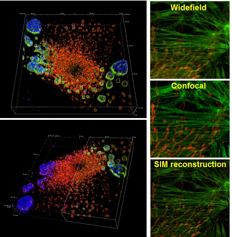
Our new Structured Illumination super-resolution Microscope (SIM) delivers twice the resolution of traditional diffraction limited microscopes (~115 nm lateral and ~300 nm axial) Capabilities: SIM enables detailed visualization of intracellular structures and their functions in fixed and live cells and tissues. 3D SIM improves lateral resolution to 115 nm and axial resolution to 269 nm and has the capability of optical sectioning of specimens, enabling the more detailed visualization of cell structures at higher spatial resolutions.
Features:
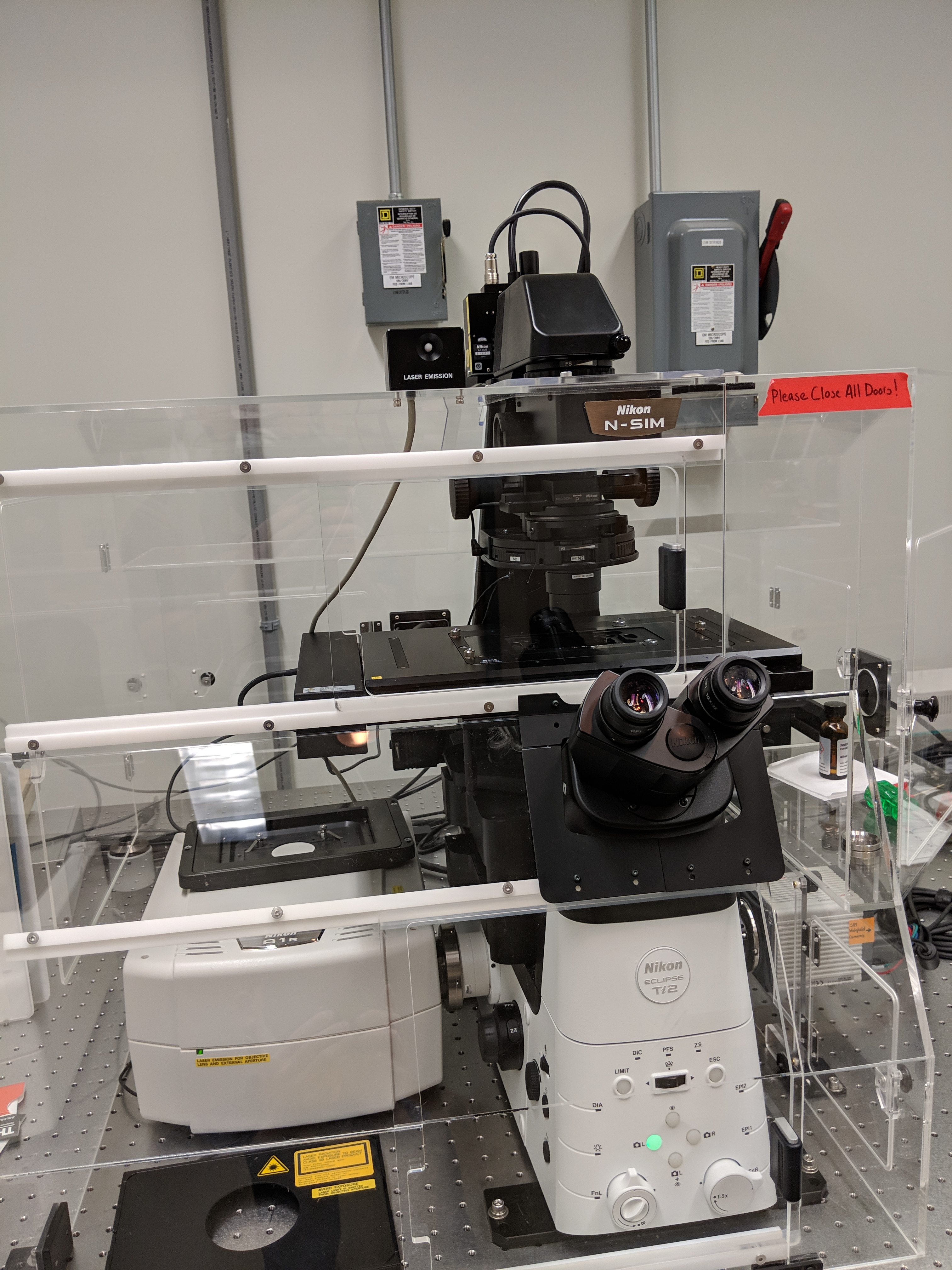
- Nikon Ti2 eclipse inverted microscope
- Prior Scientific motorized XY stage
- Multiple region time lapse with z-stack for 3D imaging. Timelapse high-speed super-resolution imaging at 15 fps.
- Nikon Perfect Focus
- Photo-manipulation (photo-bleaching/photo-activation) experiments can be easily conducted with multiple user-defined regions of interest
- Advanced Detection: Hamumatsu Orca Flash 4 Digital Camera
- Automatic switching between illumination modes: The newly-developed high-speed structured illumination technology enables fast acquisition rates, automatic switching between illumination modes, and automated optimization of structured illumination patterns for different wavelengths and magnifications. This expanded automation enables fast 2-color TIRF-SIM imaging, as well as multiplexing of different SIM modalities. The N-SIM S provides easy-to-use, streamlined workflows, whether it be for single-mode or multi-modal imaging experiments.
- TIRF-SIM/2D-SIM mode: This mode captures super-resolution 2D images at high speed with incredible contrast. The TIRF-SIM mode enables Total Internal Reflection Fluorescence observation at double the resolution of conventional TIRF microscopes, facilitating a greater understanding of molecular interactions at the cell surface.
- 3D-SIM mode: Generates structured illumination patterns in three dimensions to deliver a two-fold improvement in lateral and axial resolutions. Two reconstruction methods (“slice” and “stack”) are available to optimize results according to application requirements (e.g. sample thickness, speed, etc.). Slice reconstruction is suitable for imaging living cells at specific depths, as it allows axial super-resolution imaging with optical sectioning at 300 nm resolution. Stack reconstruction, based on Gustafsson’s theory, is suitable for acquisition of volume data as it can image thicker specimens with higher contrast than slice reconstruction.
Objectives:
- Plan Apo λ 4x na . 2 wd 20000
- Plan Apo λ 10x na . 5 wd 4000
- Plan Apo λ 20x na .8 wd 1000
- Plan Fluor 40x DIC h n2 na 1.3 we 240um
- Apo 60x λ oil DIC n2 na 1.4 wd 140
- SR Apo Tirf 100x na 1.5 wd 120
Laser Lines(nm):
405, 488, 567, 637
Filter Cubes(eyes):
DAPI, FITC, TRITC
Accessories:
- Tokai Hit incubation stage chamber at 37ºC, optional 5% CO2
- Motorized x-y stage
- Z-stacks
- Nikon Perfect Focus System
Acquisition Software:
The Nis Elements 5.0 Imaging Software contains a cutting-edge analysis package of modules for doing quantification and densitometry of fluorescence in 2D and 3D. Nis Elements can also be used to count cells and track cells on time-lapse movies.