 |
2013 Case #5 |
 |
| The following patient was seen in the outpatient department of the 36-bed Tropical Disease Unit at Cayetano Heredia National Hospital. |
|
Epidemiology: Born and lives in the jungle of Pichanaki; works as a farmer harvesting coffee and oranges. No known TB exposure; no risk factors for STDs or HIV. Physical Examination: Afebrile. Edema and infiltration of the lower and upper lips; several painful oral lesions that bleed easily on examination; infiltration of the gingiva; poor dentition [Images A & B]. Examination of the hard and soft palate and larynx were normal. Chest clear to auscultation. Cervical lymphadenopathy; lymph nodes were small, around 1cm, non-tender, non-fluctuating. Laboratory Results: Hb 12.1 g/dl; WBC 6500 (0 eos, 60% neutrophils, 30 lymphs, 10 monos). Normal glucose; normal hepatic and renal function. Stool O&P negative. Negative HIV and HTLV-1. Chest x-ray is shown in Image C. |
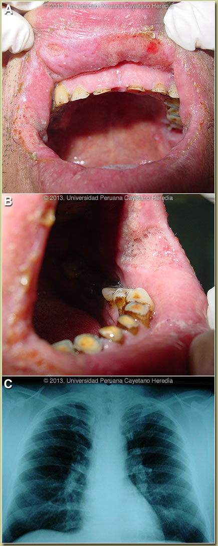
| Diagnosis: Paracoccidioides brasiliensis infection (chronic form) with mucosal involvement. |
|
The major differential diagnosis in Perú of oro-pharyngeal lesions in non-HIV infected patients would be mucosal leishmaniasis, paracoccidioidomycosis, carcinoma, or lymphoma. In Perú, leishmaniasis with destructive but painless lesions would be by far the most common. In general, oral lesions of paracoccidioidomycosis are painful, are frequently friable and bleed on contact, and gingival and buccal mucosa are frequently involved. Transmission is primarily by inhalation. Chest x-ray is typically abnormal [see Gorgas Case 2012-07 discussion). However, there may be patients with no symptoms and abnormal chest x-rays or patients, like this case, with no symptoms and normal x-rays. Autopsy studies have shown that almost all patients have pulmonary abnormalities in the absence of abnormal x-rays, reflecting that the primary lesion is in the lungs. When CT scans are performed, abnormalities are usually observed, even at early stages of infection. The differential diagnosis for the lung disease with an abnormal chest x-ray includes: TB, histoplasmosis, lymphoma, cancer and cryptococcosis. The most typical radiographic pattern of paracoccidioidomycosis is with bilateral mixed infiltrates (alveolar, interstitial, and nodular), mainly located in the middle and lower lobes. Interstitial lesions may have a miliary, nodular or fibronodular pattern. Other patterns observed in these patients are hilar and mediastinal lymph node enlargement, cavities, and calcified lesions. Extra-pulmonary disease is found in over 70% of cases and may involve skin, mucous membranes, lymph nodes, adrenals, abdominal organs and CNS (in 10%). Bacterial superinfection of ulcerative oral lesions when they occur is more common than with oral ulcers due to mucocutaneous leishmaniasis. Paracoccidioidomycosis is most often an indolent and relapsing disease. The chronic form (adult type) of the disease is believed to represent reactivation of latent infection initially acquired via inhalation. The chronic forms represent approximately 95% of all cases in the experience at our institute (120 patients through 2010), and approximately 85% in the Brazilian series [Rev Soc Bras Med Trop. 2003 Jul-Aug;36(4):455-9]. See examples of this form in previous Gorgas Cases 2005-12, 2009-06, and 2004-05. We have also previously shown a case of the chronic progressive form of the disease [Gorgas Case 2003-07] and the relatively uncommon “juvenile” acute progressive and disseminated form of the disease, which usually (but not always) affects adolescents and young adults [Gorgas Case 2010-07]. TB coexists in up to 10% of patients with paracoccidioidomycosis. Cavitation and pleural effusion are less commonly seen than in TB. Paracoccidioidomycosis, also known as South American blastomycosis, is found in humid forested or lush green areas of the Americas from Southern Mexico south to Uruguay and Argentina. It appears to be most common in Brazil. The exact habitat of the organism is unclear but transmission is described as being entirely by airborne inhalation. However, we have observed cases with only oral lesions apparently associated with the use of tree leaves contaminated with fungal spores as toothpicks. Primary pulmonary infection may be asymptomatic and self-limited, but even with treatment, will produce at least moderate pulmonary fibrosis. Rural adult male agricultural workers between 30-60 years of age are most affected by the infection. Travelers spending less than 6 months in an endemic area are unlikely to acquire paracoccidioidomycosis. Sulfonamides, ketoconazole, itraconazole, and amphotericin B are all effective therapies. For typical cases, itraconazole 100-200 mg/day for 6-9 months is regarded as the treatment of choice when it is available and affordable. Relapses are common with less than 6 months of therapy and expert opinion is now that 1-year therapy is not necessary. In the developing-world setting, ketoconazole is likely equally effective and is usually less than half the cost. However, 12 months of therapy with ketoconazole is generally recommended. This patient was started on itraconazole 200mg/d with improvement. The plan is for at least 6 months of treatment. |
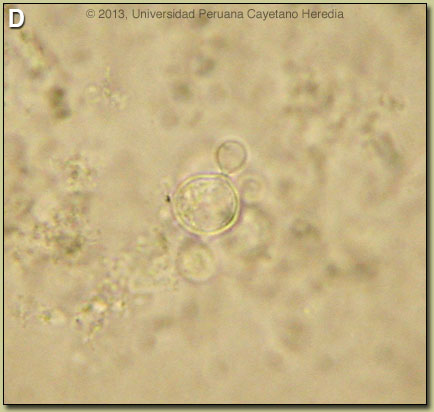 Discussion: The diagnosis was made by simple KOH preparation of a mucosal scraping, which showed a typical spherical cell, 10-40 microns in diameter with a thick birefrigent cell wall [Image D]. The yeast is surrounded by peripheral buds with typical narrow necks. When completely surrounded by such buds a so-called “pilot-wheel” pattern occurs. Direct scrapings will be positive in up to 90% of cases of paracoccidioidomycosis with oral lesions. In this case sputum AFB stain was negative. Biopsies are not necessary in characteristic clinical cases with positive scrapings.