 |
Gorgas Case 2014-03 |
 |
|
The following patient was seen in the outpatient department of the Tropical Disease Unit at Cayetano Heredia National Hospital in Lima, Perú.
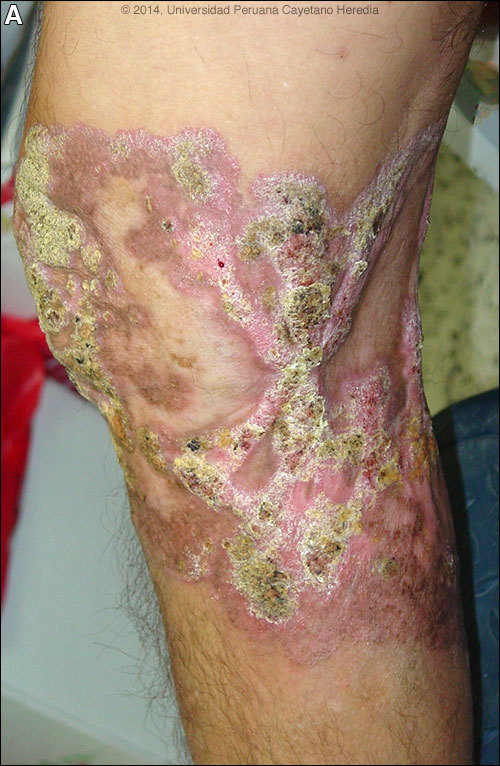 History: 41-year-old male with a 21-year history of slowly progressive verrucous lesions around the right knee. Initial painless pruritic papules localized to the medial part of the knee spread gradually over 1 year and all became verrucous and in addition to pruritus, a burning sensation was also noted. No history of any specific trauma. No drainage at any time. Since then, the lesions have progressed very slowly. No other skin lesions at any time. No fever or constitutional symptoms. Many courses of antibiotics and topical creams resulted in no improvement. Some difficulty in extending the knee has developed in the last 2 years. He was presented during a visit to another city to one of our physicians who referred him to us. Epidemiology: Lives and grew up in a rural city of Contamana, along the Ucayali River in the Amazon of Perú. Works as a farmer harvesting corn, with exposure to fresh water lakes and ponds, mostly with bare feet. No HIV risk factors. Physical Examination: Afebrile; normal vital signs. No lymphadenopathy. Extensive involvement of the entire right knee with plaques over the anterior and posterior aspects [Image A]. The lesions have a verrucous, elevated border, with areas of central scarring. No lymphedema, no suppuration. Chest: clear. Abdomen: no hepatosplenomegaly. Neurological: normal. Laboratory Results: Hb 12.4; WBC 6800 (68 neutrophils; no eos). Normal biochemistry and urine. Blastocystis hominis was found in the stool. Normal chest x-ray and normal plain films of both legs and knees. Negative serological tests for HB, HTLV-1 and HIV.
|
|
Diagnosis: Chromoblastomycosis.
Acknowledgement: We would like to thank Dr. Francisco Bravo, Gorgas Dermatology Professor and Dr. Betty Bustamante, Director of the IMT Mycology Lab. 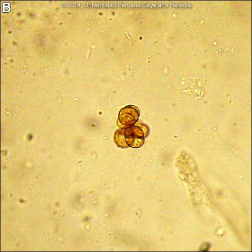 Discussion: A 10% KOH preparation of a skin scraping of a verrucous lesion [Image B] disclosed pigmented thick-walled multi-celled structures called sclerotic bodies, medlar bodies, or muriform bodies, which are diagnostic of the clinical syndrome of chromoblastomycosis. These are double-walled brown structures with a diameter of 4 to 10 μm that resemble copper pennies. Thick and dark hyphae are sometimes identified. Cultures can be performed on Sabouraud agar or Sabouraud with antibiotics at 25°–28°C. The organism grows slowly (25-30 days). Culture and pathology are pending. Subcutaneous mycoses are a heterogeneous group of diseases and organisms that share the characteristic that they develop at the site of skin penetrating trauma. Etiologic agents of subcutaneous mycoses are divided into several clinical groups:
Chromoblastomycosis occurs worldwide, including in the USA, but 70% of cases are estimated to occur in the moist tropics. Most cases in Latin America are from the humid Amazon of Brazil, from Mexico and from Costa Rica, but cases are reported from Colombia, Ecuador, Venezuela, Argentina, the Dominican Republic and Cuba. Many cases are reported from Madagascar and South Africa. Up to 90% of cases occur in males likely due to occupational factors. The differential diagnosis of a life-long subcutaneous indolent infection like this includes: sporotrichosis [Gorgas Case 2008-01], eumycetoma [Gorgas Case 2002-04] (no drainage with grains were reported in this case), lobomycosis (lacaziosis) [Gorgas Case 2005-03], subcutaneous zygomycosis, subcutaneous phaeohyphomycosis, botryomycosis [Gorgas Case 2003-04], leishmaniasis [Gorgas Case 2004-01], nocardiosis [Gorgas Case 2011-01], and tuberculosis verrucosa cutis. Squamous cell carcinoma and leprosy are less likely. Our patient is classified in the moderate group of illness based on an old classification system that takes into account four criteria: total area of involvement, number of lesions, complications, and lack of response to prior treatment. Long-term complications include superinfections, ulceration and lymphedema. Treatment of chromoblastomycosis is often difficult and is based on case reports and case series; there are no controlled trials. Treatment choices include itraconazole alone or in combination with flucytosine or terbinafine (expensive and not typically available in the tropics). With very chronic infection like this, itraconazole may induce significant fibrosis that impairs functionality of the extremity. Duration of treatment is no less than 1 year starting at 200mg tid for 3 days, then 200mg bid. Measurement of serum drug levels is recommended to improve outcomes. Response is observed after 3-4 months of therapy; sustained response is not always observed due to its fungistatic nature. Of the newer azoles, voriconazole has potent in vitro activity against Cladophialophora carrionii, F. pedrosoi, and P. verrucosa; posaconazole is approved in Europe for refractory chromoblastomycosis; and caspofungin has potent in vitro activity against F. pedrosoi [Med Mycol. 2011 Apr;49(3):225-36].
|