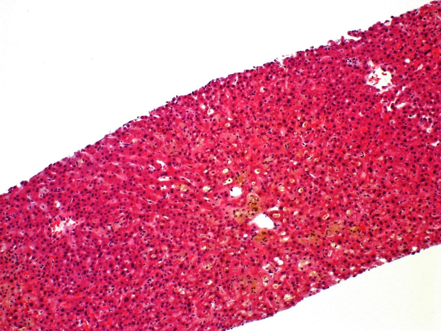
1) A 45-year-old woman described increased fatigue over a period of 1 week and decided to get a doctor’s appointment after her spouse noted scleral icterus. Family history is notable for a maternal aunt with PBC. Her medical history was notable for cholecystectomy 5 months ago and detection of hookworm infection (treated with thiabendazole) 3 weeks prior to this clinic visit. Laboratory studies revealed elevated transaminases and total bilirubin. Liver biopsy revealed bland cholestasis with minimal mixed portal infiltrates. No ductular reaction or granulomatous inflammation were seen. What is her underlying condition?
Choose the Correct Diagnosis:
- Cholecystectomy related cholangiopathy
- Drug-induced liver injury
- Primary biliary cholangitis
Answer: B.) Drug-induced liver injury
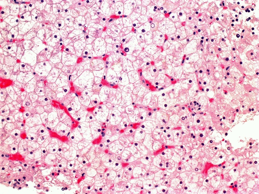
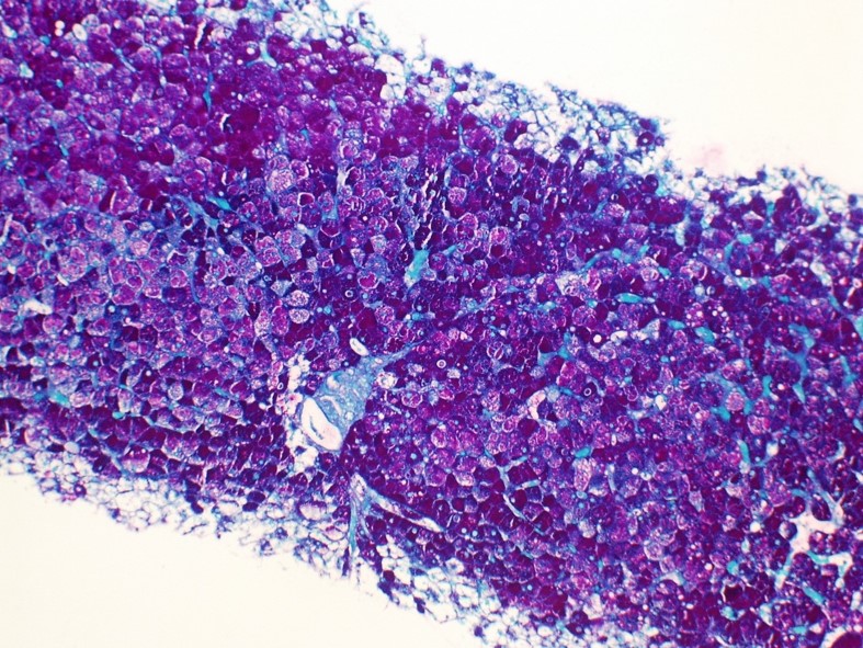
A 20-year-old man presented to the ER with symptoms of diabetic ketoacidosis. After stabilization, lab work revealed elevated liver enzymes. Liver biopsy was performed. What is his condition?
Choose the Correct Diagnosis:
- Alpha-1 antitrypsin storage disorder
- Severe steatohepatitis
- Glycogenic hepatopathy
Answer: C.) Glycogenic hepatopathy
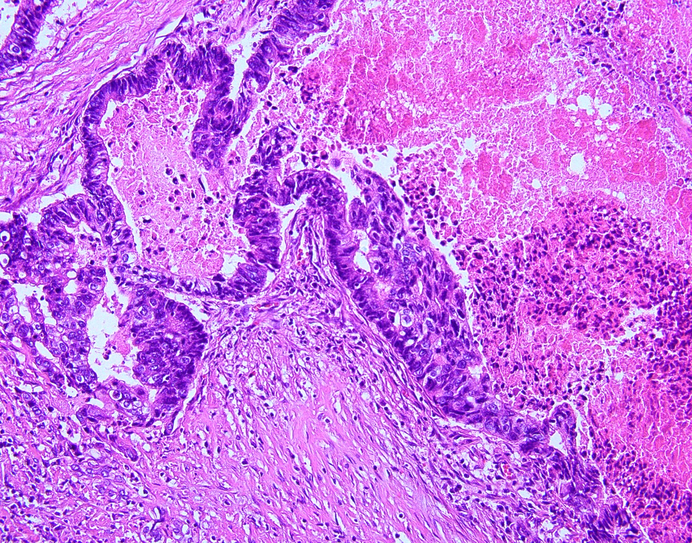
A 60-year-old woman was noted to have severe anemia and symptoms of colonic obstruction. Abdominal CT revealed a large lesion in a non-cirrhotic liver. Immunostains performed on a hepatic biopsy specimen revealed lesional cells to be positive for CK20 and CDX2 and negative for CK7, glypican-3, and TTF-1.
What is the lesion?
- Metastatic colonic adenocarcinoma
- Cholangiocarcinoma
- Metastatic mammary carcinoma
Answer: A.) Metastatic colonic adenocarcinoma
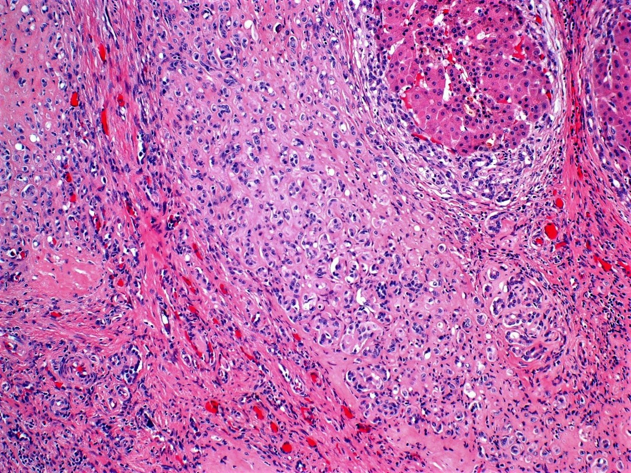
A 21-year-old man presented with a clinical history of cryptogenic cirrhosis. Grossly, the explant liver was noted to be very firm. Sectioning revealed parenchymal color varying from dark red to tan white with dilated ducts and apparent stones. No discrete lesion was identified. Histologic examination revealed numerous partially involuted hepatic regenerative nodules often surrounded by swaths of gland-like tissue, the lumens of some showing calcifications and occasional red blood cells. Cytologically, no increased mitoses or necrosis were noted. Immunostains showed these areas to be positive for CD34, CD31, and negative for CDX2 and CA 19.9.
What chromosomal translocation is this tumor associated with?
- t(8;14)(q24;q32) IGH/MYC
- t(1;3)(p36.3;q25), CAMTA1- WWTR1
- t(11;22)(q24;q12) EWS -FLI1
Answer: B.) t(1;3)(p36.3;q25), CAMTA1- WWTR1
Contributed by Leona Council, M.D.