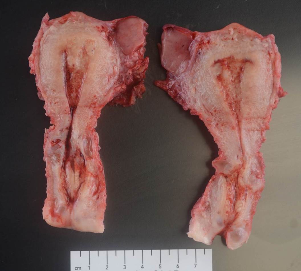
 Tumor is grossly involving uterine serosa
 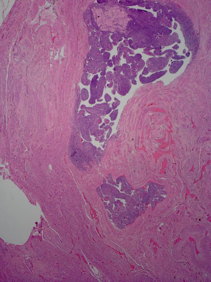 Myometrium with lymphovascular tumor invasion
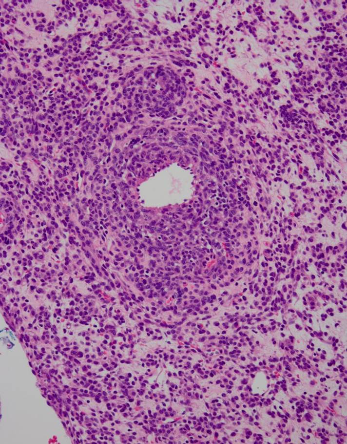 Detail of tumor with cellular perivascular areas
Clinical history:
49 year-old-female with pelvic mass and diffuse pulmonary lesions
Gross examination:
Tumor is present in the uterus and surrounding tissue (“pelvic mass”), total size 19.0 cm
Microscopic examination:
Tumor within myometrium with stromal and lymphovascular invasion and in “pelvic mass”. No mitoses are identified.
Immunohistochemical stains:
-Positive: ER, PR, SMA, desmin, HMB-45, MiTF
-Negative: CD10, CK LMW (CAM 5.2), CK HMW (34BE12), DOG1.
Choose the correct diagnosis:
- Endometrial stromal sarcoma
- Perivascular epithelioid cell neoplasm
- Intravascular leiomyomatosis
- Gastrointestinal stromal tumor
Answer: “B.” Perivascular epithelioid cell neoplasm (PEComa)
Discussion:
Age: Reproductive age and postmenopausal women
Clinical features: Uterine bleeding. Associated with lymphangioleiomyomatosis and tuberous sclerosis
Histology: Cells with well defined borders and abundant clear cytoplasm or spindle cells. Cells may be tightly arranged around blood vessels. Low mitotic activity. Occasional stromal hyalinization
IHC: Positive for HMB-45, MiTF; variably positive for SMA, CD10; negative for cytokeratin
Behavior/prognosis:
Benign, borderline,malignant.
Aggressive behavior: >5 cm, infiltrative growth pattern, high-grade cytologic atypia, necrosis, mitotic activity 1/50HPF.
Presented case has an aggressive behavior based on the large tumor size, LVI and diffuse lesions in the lungs
Literature:
Perivascular Epithelioid Cell Neoplasm (PEComa) of the Gynecologic Tract. Clinicopathologic and Immunohistochemical Characterization of 16 Cases
John Kenneth Schoolmeester, MD, Brooke E. Howitt, MD, Michelle S. Hirsch, MD, PhD, Paola Dal Cin, PhD, Bradley J. Quade, MD, PhD,w and Marisa R. Nucci, MD. Am J Surg Pathol 2014;38:176-188
Case contributed by Lea Novak, M.D., Associate Professor, Anatomic Pathology