Case history:
40-year-old male with rapidly growing thyroid tumor.
Diagnosis:
- Angiosarcoma
- Follicular thyroid carcinoma
- Anaplastic thyroid carcinoma
- Medullary carcinoma
Answer:
“C”--Anaplastic thyroid carcinoma
Discussion:
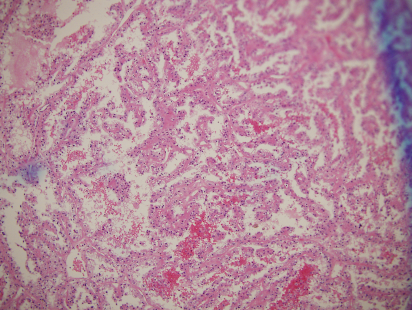
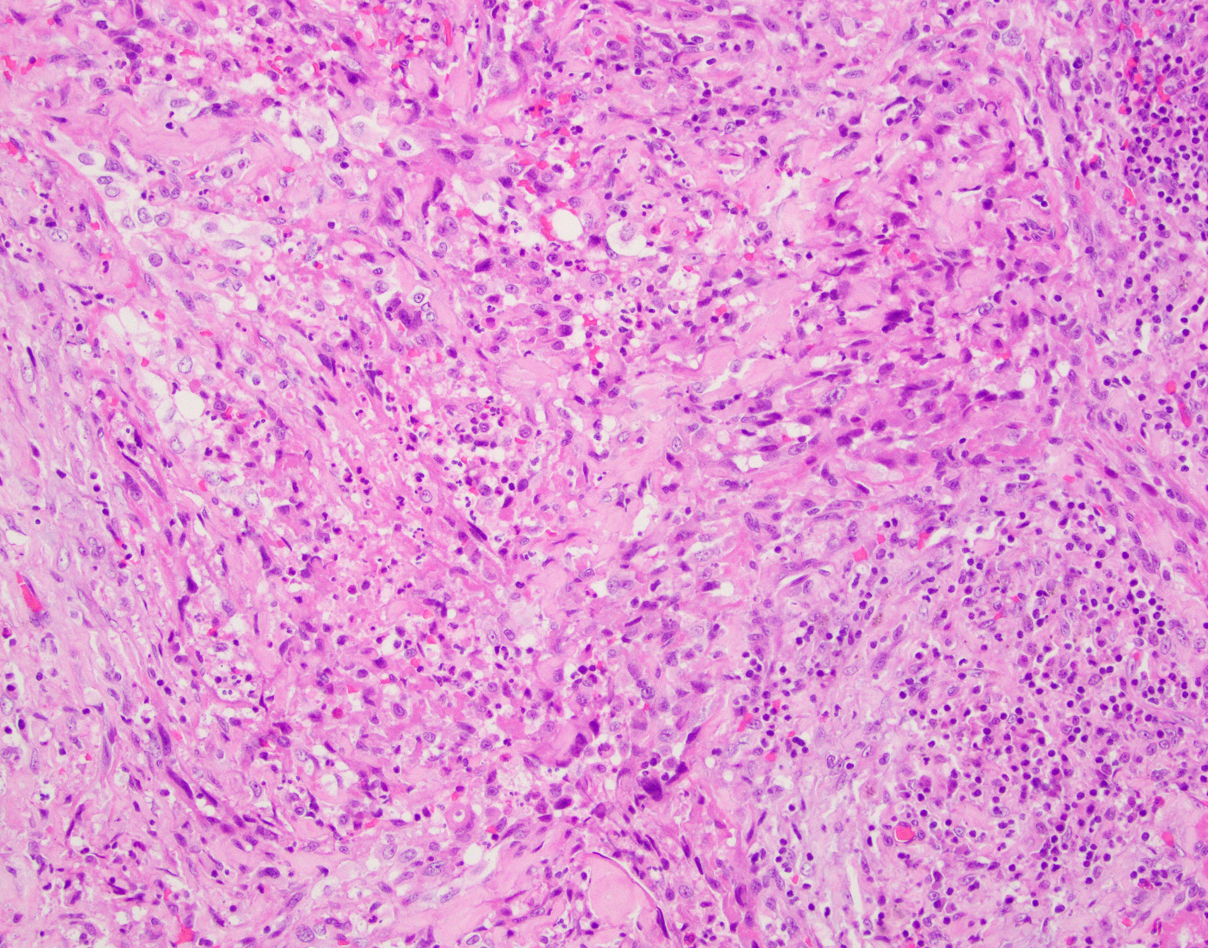
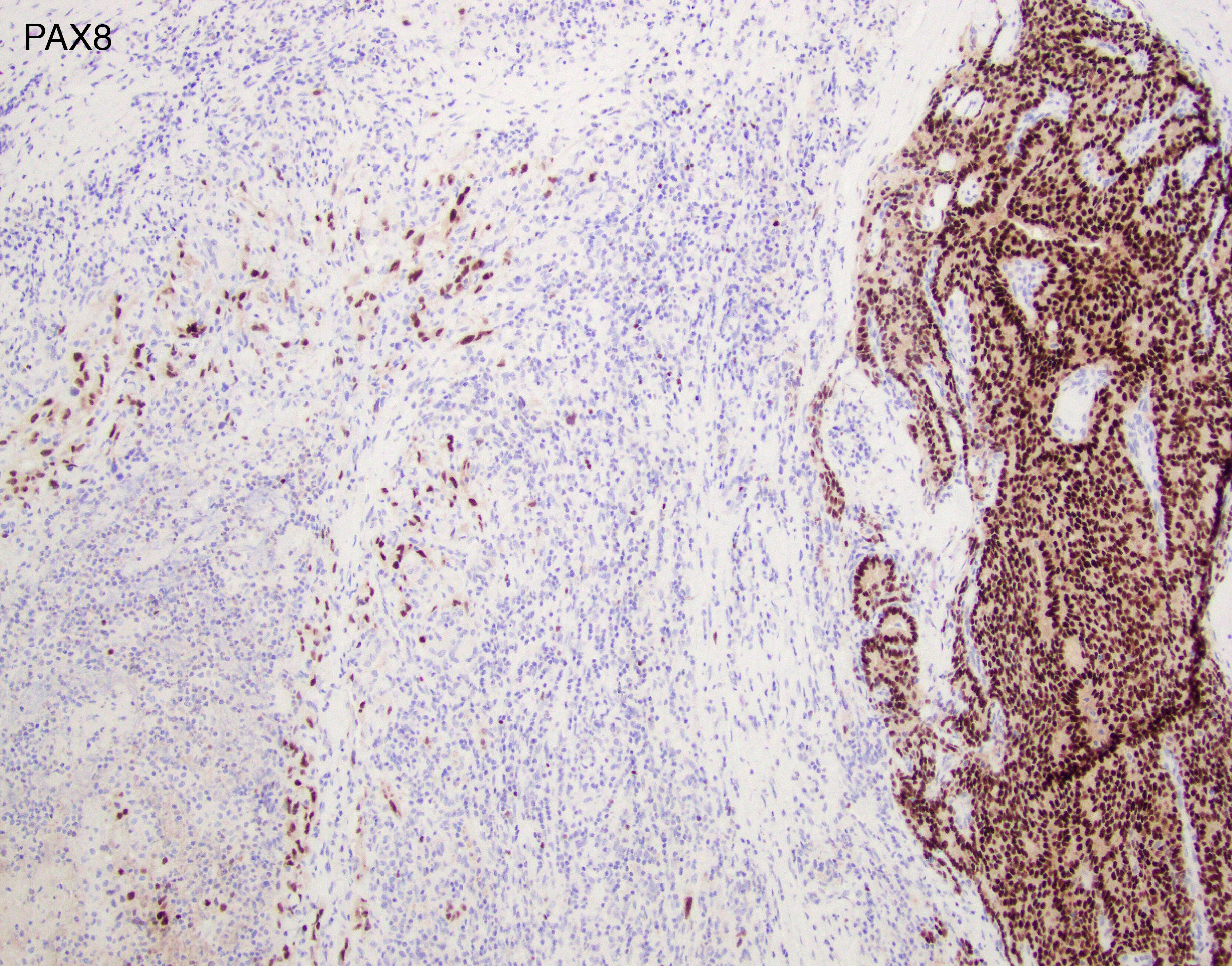
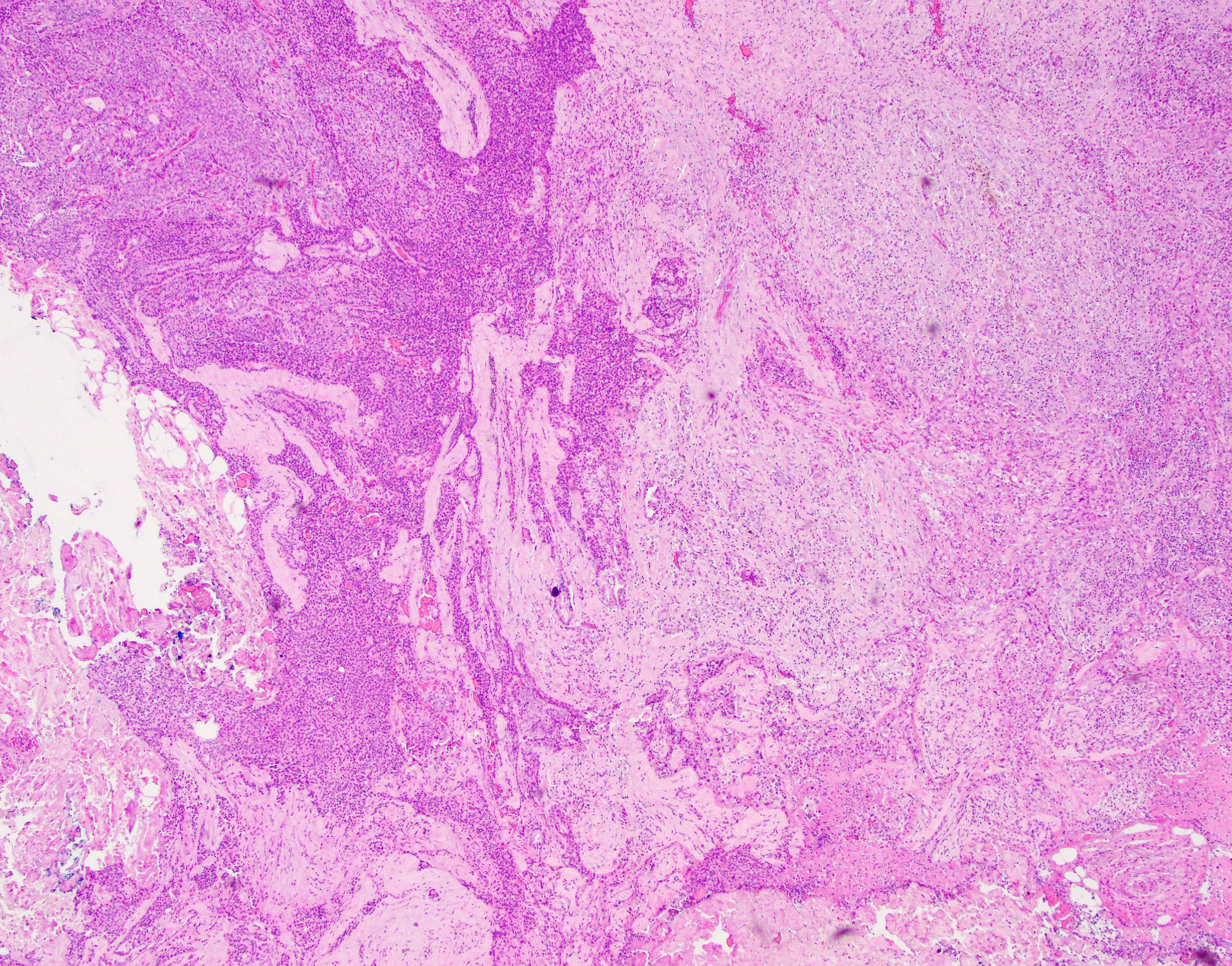
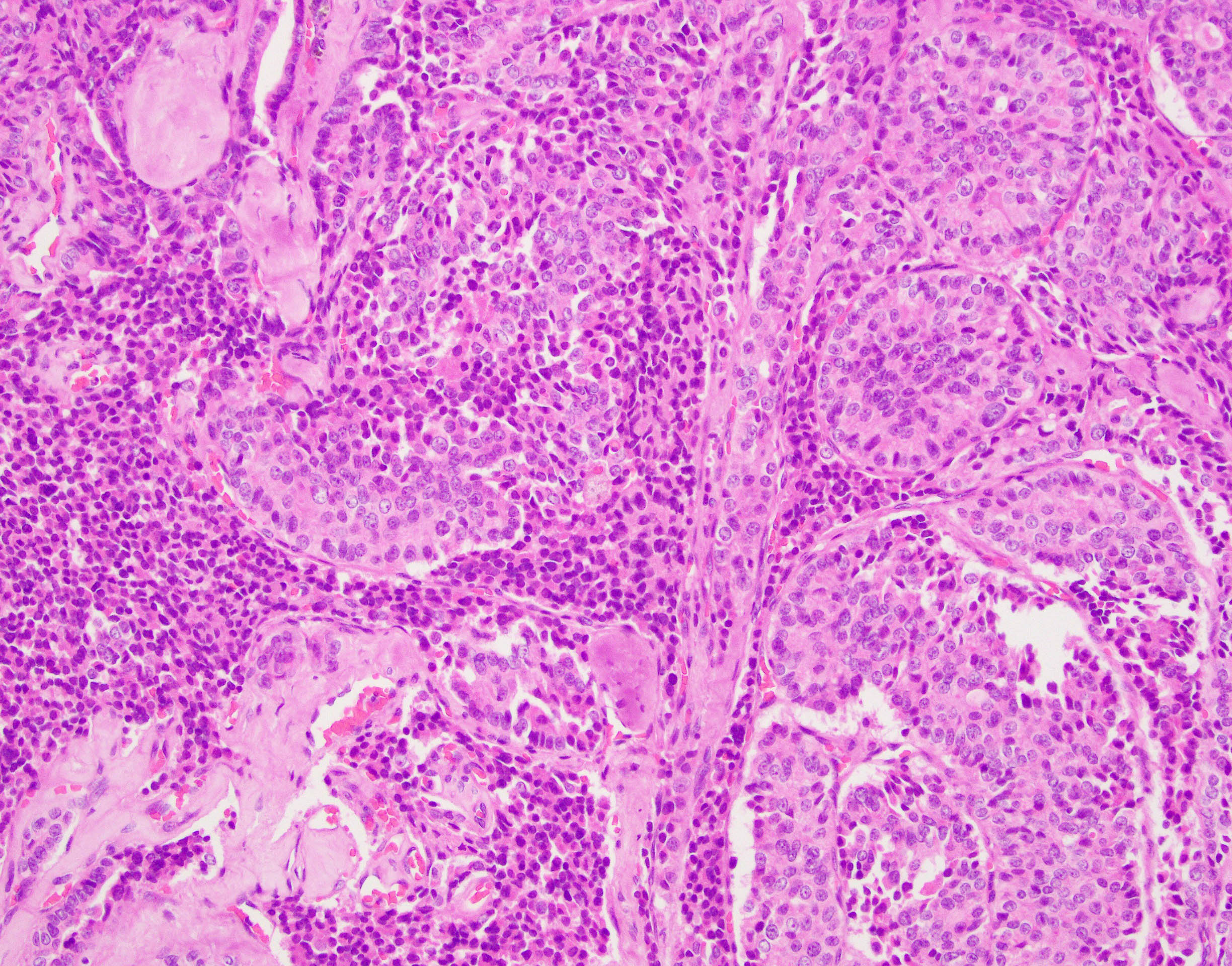
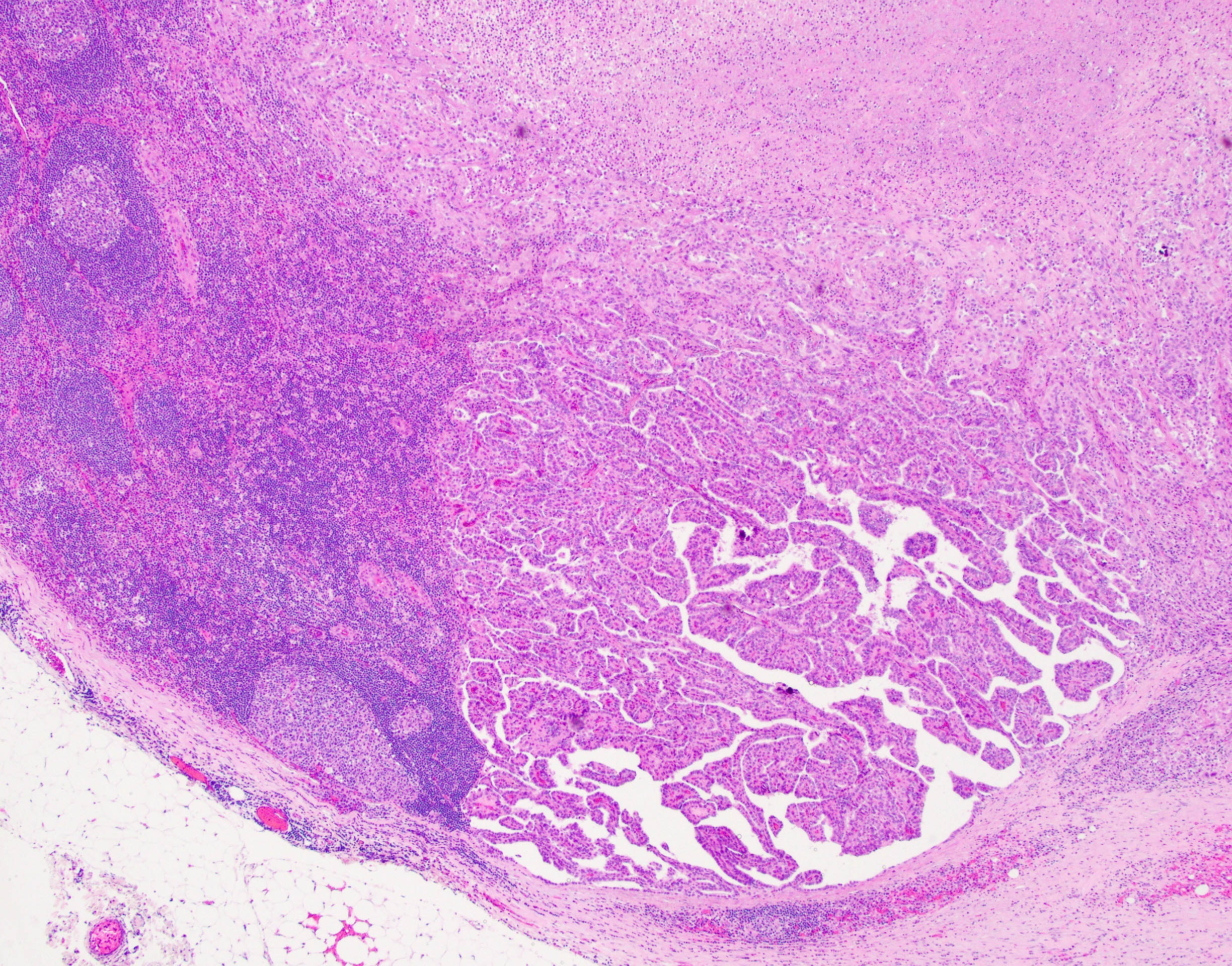
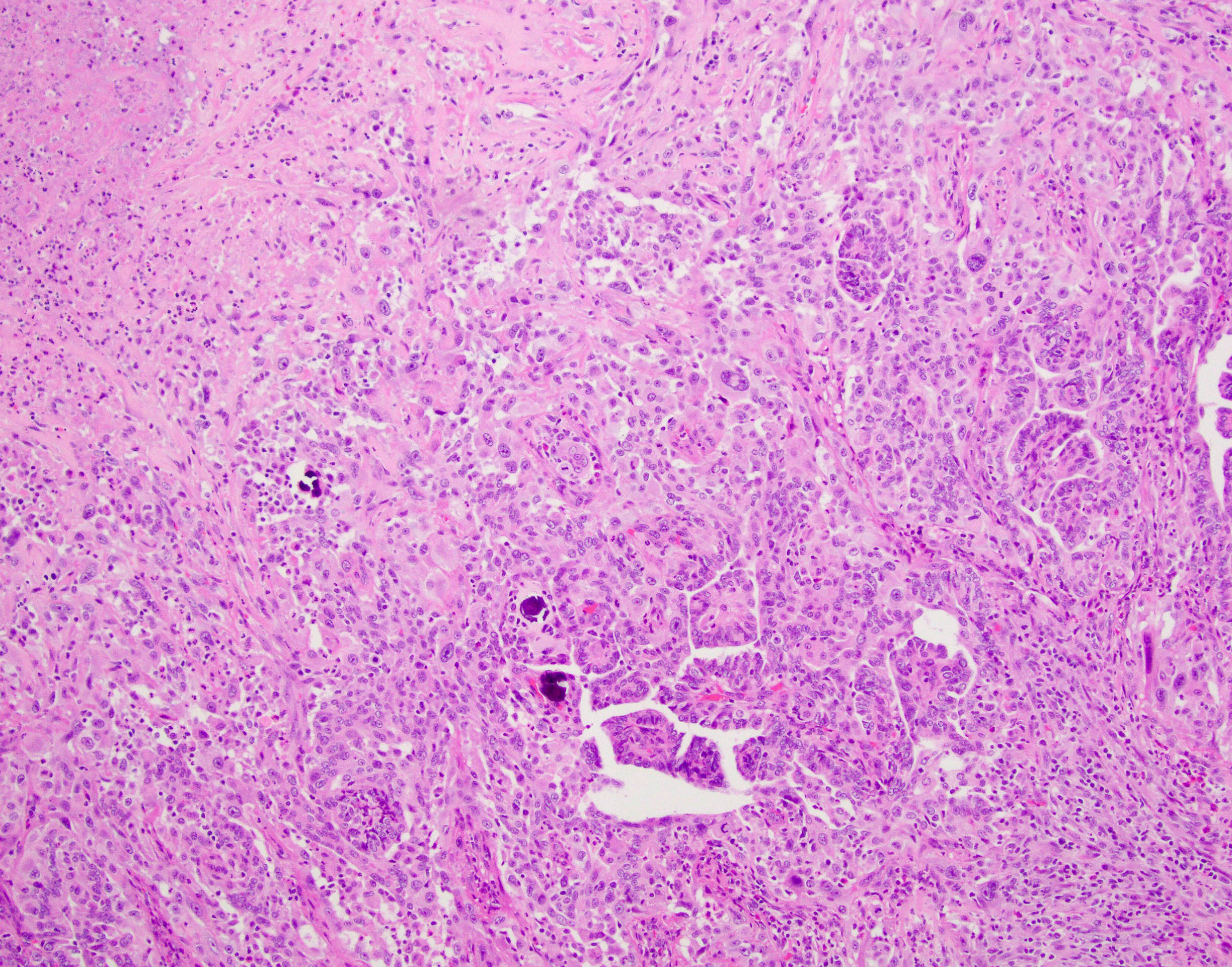
This dramatic example of anaplastic carcinoma illustrates the different steps involved in its carcinogenesis. On the right hand side of figure 1 one can appreciate the precursor lesion of papillary thyroid carcinoma (PTC) with a sharp transition to the anaplastic component (left side of picture) that is made of sheets of highly malignant squamoid and sarcomatoid cells punctuated by patchy areas of geographic necrosis. Immunohistochemical stains for TTF1 and thyroglobulin (not shown) highlighted only the PTC component but immunoreactivity for these markers was completely lost in the anaplastic component. However, PAX8 (figure 3) and Cytokeratin expression (not shown) was retained in both components highlighted their usefulness when dealing with anaplastic thyroid carcinoma. Interestingly, the precursor PTC component exhibited some areas of insular–like growth pattern with lack of PTC nuclear features, as seen in the so-called poorly differentiated thyroid carcinoma (figures 4 and 5); thus, displaying the progression from PTC to poorly differentiated thyroid carcinoma to finally anaplastic carcinoma. Since anaplastic carcinomas are extremely aggressive tumors that tend to obliterate everything in their path, including potential precursor lesions, observation of this stepwise tumor progression on a specimen is somewhat rare. Another interesting finding in this case was that the lymph node metastases also displayed tumor progression to anaplastic carcinoma (figures 6 and 7). The excellent chapter in Dr. CDM Fletcher’s book on thyroid and parathyroid tumors written by Dr. John K. C. Chan contains a graphic illustrating the relationship between these tumor types
(Chapter 18, pp. 1219, Figure 18A-76).
References:
Diagnostic Histopathology of Tumors Vol. 2, 4th Edition, Fletcher CDM ed. Elsevier
Case contributed by Carlos Prieto-Granada, M.D., Assistant Professor, Anatomic Pathology, UAB Department of Pathology