Case history
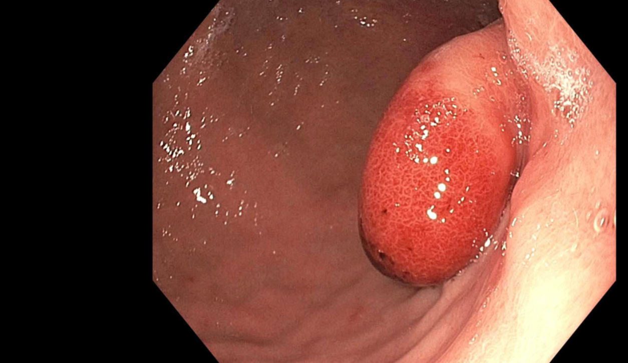
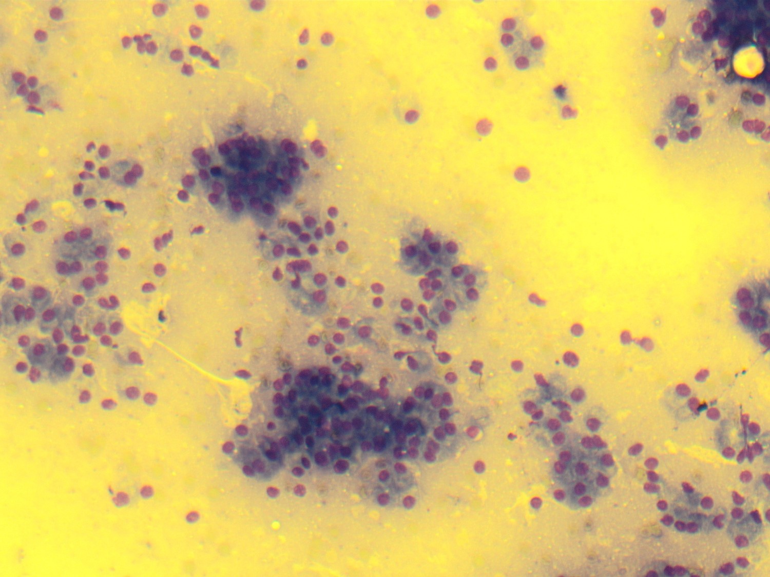
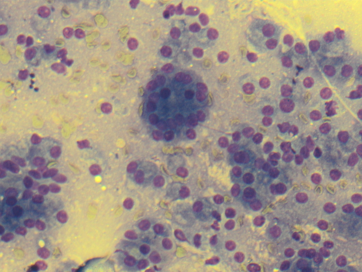
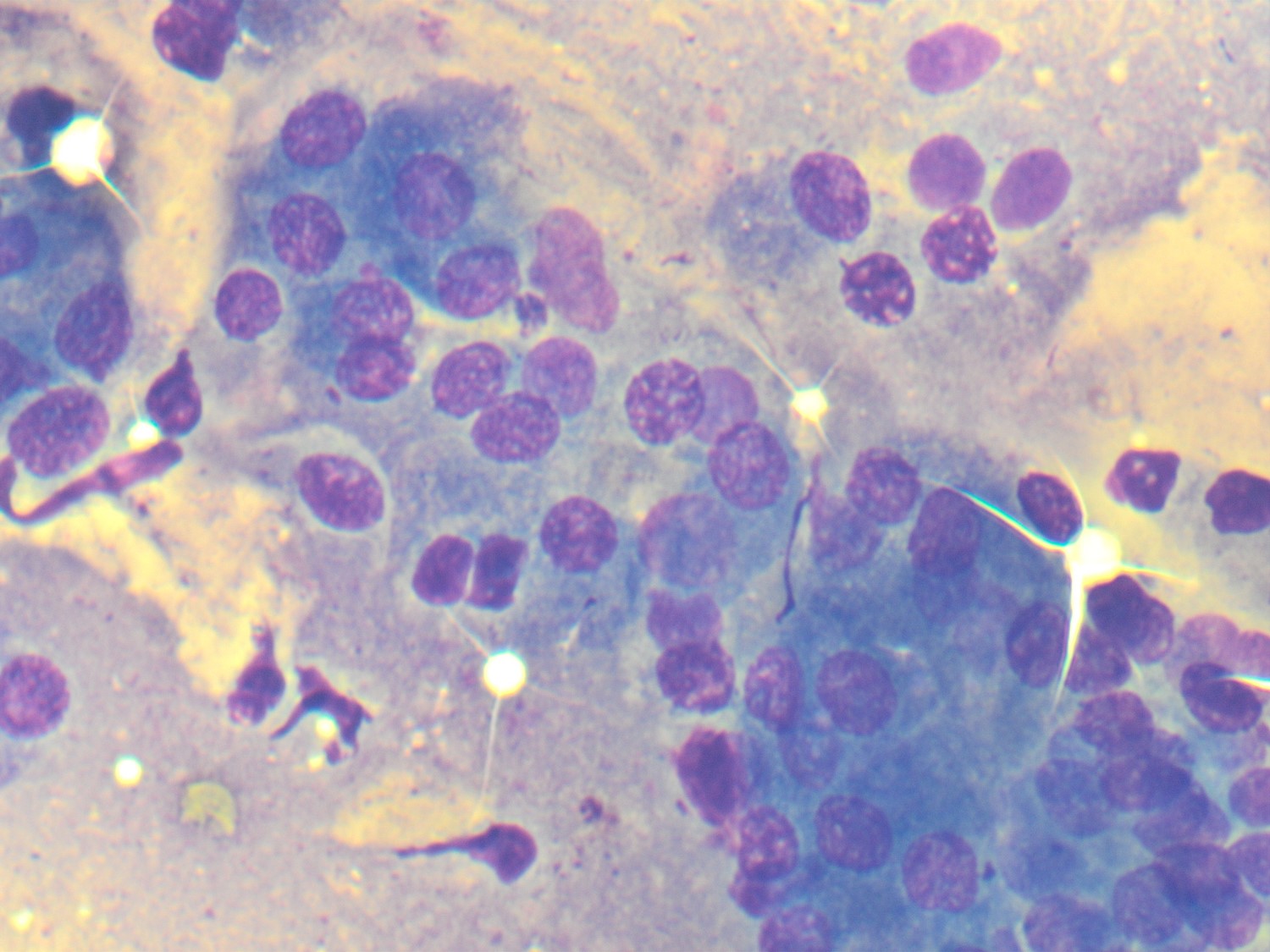
A 36 year old man presented to an OSH for an upper GI bleed. Biopsy was non diagnostic. He was referred here for EUS-FNA. US showed an 11 mm. hypoechoic submucosal mass in the lesser curvature (Fig 1). EUS-FNA smear stained with Diff-Quik is moderately cellular (Fig 2-4).
Diagnosis:
- Gastric adenocarcinoma
- Endocrine neoplasm
- Pancreatic heterotopia
- Epithelioid GIST
Answer:
The correct diagnosis is "C. Pancreatic heterotopia."
Discussion:
On FNA, coarsely granular cytoplasm, acinar formation, and uniform round nuclei with low N:C ratios suggested the final diagnosis, although an endocrine neoplasm could not be excluded. IHC stains of the cell block were inconclusive due to insufficient lesional material.
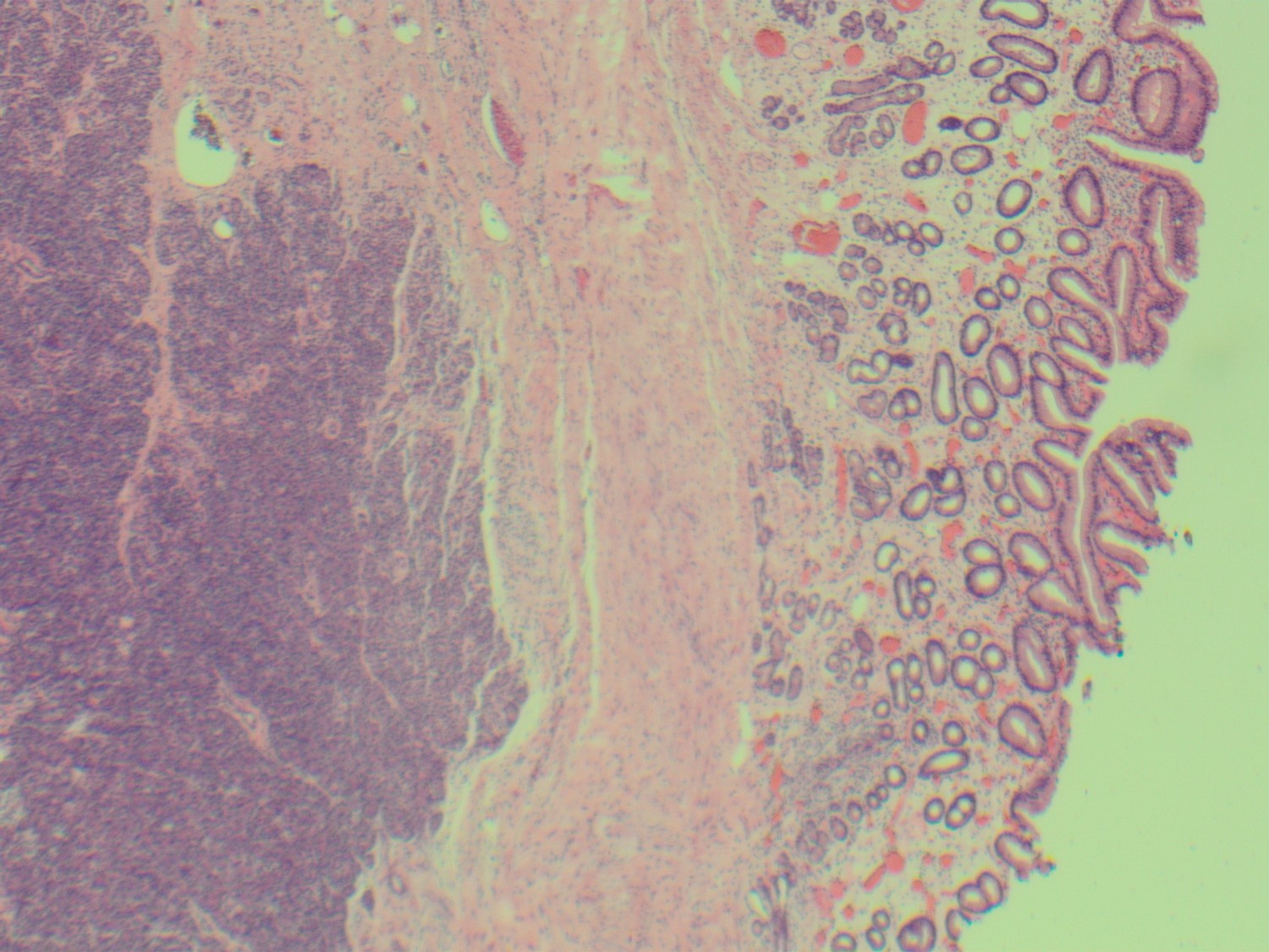
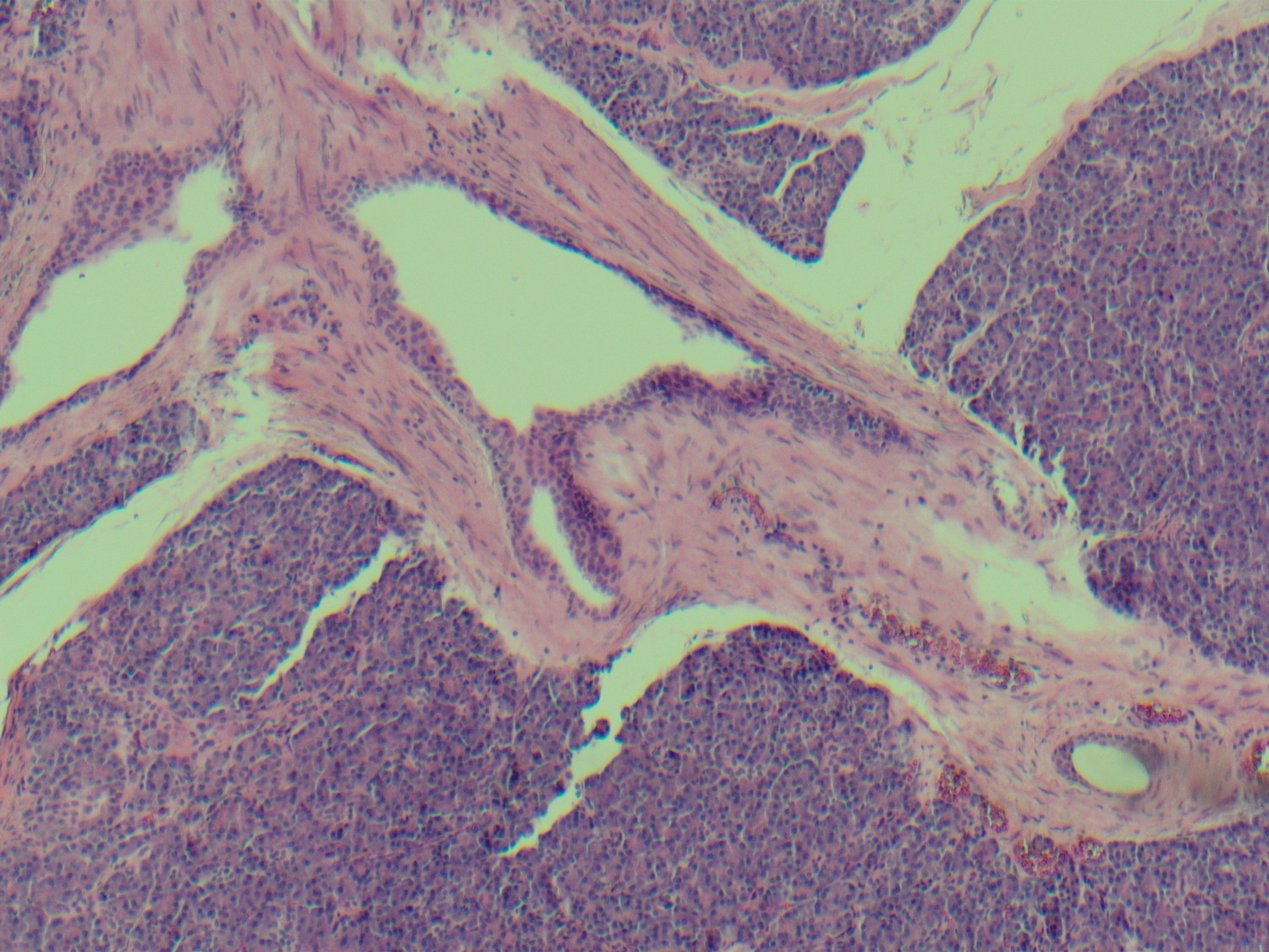
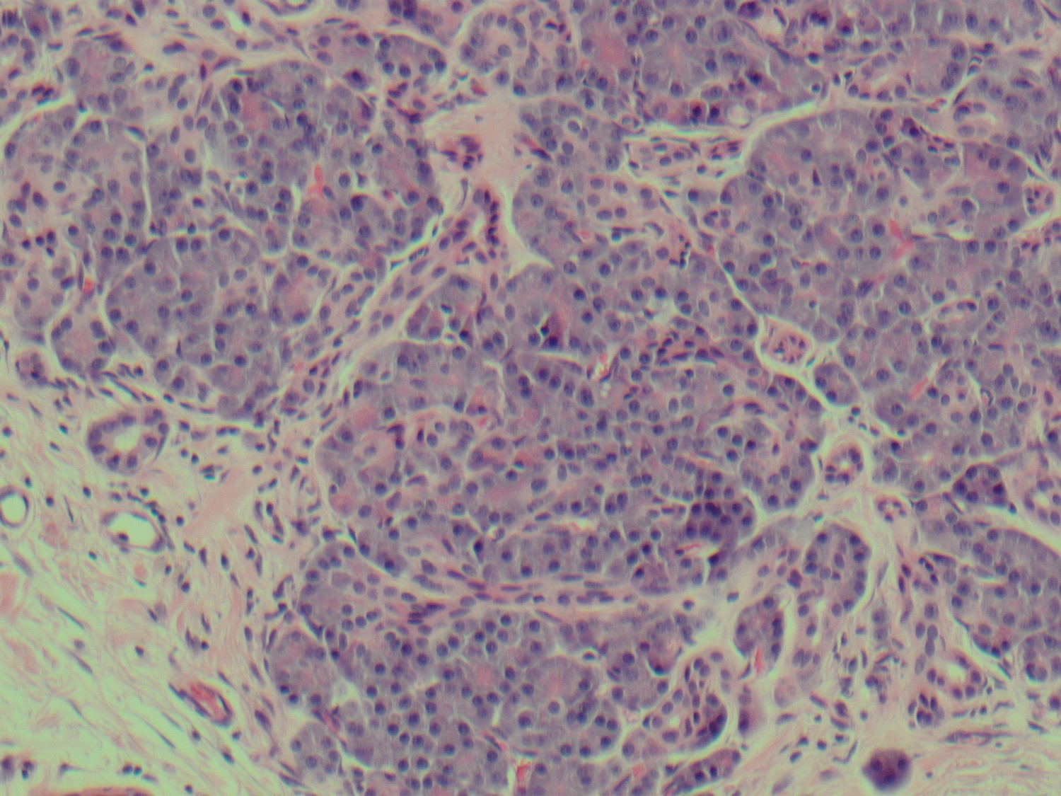
The lesion was resected, showing a submucosal rest of histologically normal pancreatic parenchyma (Fig 5-7).
In some series, heterotopic pancreas comprises up to 4% of benign gastric polyps. Men and women are affected equally. The polyps can occur at any age and are usually less than 3 cm. in diameter.
Case contributed by Ralph Crowe, M.D., Professor, Anatomic Pathology, UAB Department of Pathology