Case History
53-year-old female with uterine mass.
What is the diagnosis?
A. Endometrial stromal sarcoma, low grade
B. Epithelioid leiomyoma
C. Adenosarcoma
D. PEComa
Answer: D
PEComa is a tumor thought to be derived from perivascular epithelioid cells. PEComas shows evidence of melanocytic and smooth muscle differentiation. PEComas affects a wide age range of patients and may arise in many locations including retroperitoneum, abdomen, pelvis, GYN organs, kidney, liver, etc. When occurring in lung it is known as clear cell “sugar” tumor or lymphangioleiomyomatosis.
In the uterus, most tumors occur in perimenopausal women; some are seen in patients with tuberous sclerosis. Tumors can be small or large, ranging in size from 0.5 to 13 cm.
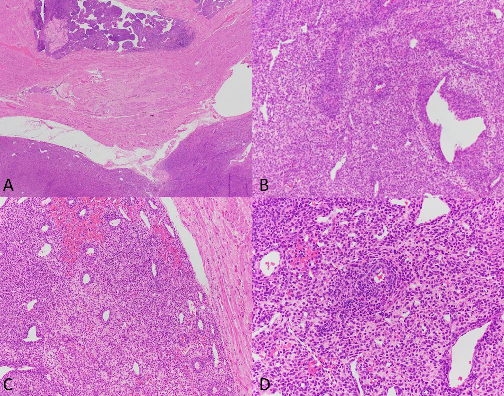
Histologically, they can be well circumscribed or infiltrative, with a ‘tongue-like’ appearance. Variable epithelioid cells, arranged in nests or diffuse sheets, to spindled cells, arranged in short fascicles, may be present. A delicate vascular network is typical and tumor cells often cluster around these vessels. Cytoplasm is eosinophilic to granular. So-called “spider cells”, formed when cells have central eosinophilic cytoplasm surrounded by a peripheral clear zone, may be present.
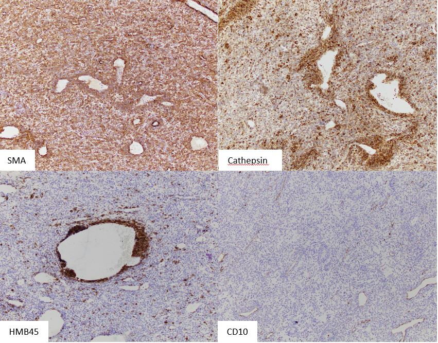
HMB45 is the most sensitive immunohistochemical marker, being expressed in up to 92% of cases. Melan-A and MITF are positive in a significant portion. About 80% stain for smooth muscle actin. A subset of cases show nuclear TFE3 expression. Genetically, PEComas may show inactivation of the TSC1 or TSC2 genes with subsequent activation of the mTOR pathway.
Criteria proposed for assessing malignant potential are tumor size > 5cm, high nuclear grade and cellularity, readily identifiable mitotic figures, infiltrative growth, necrosis and vascular invasion. Tumors with 2 or more of these high-risk features are considered malignant. Tumors 5 cm or more in size with no other high-risk features, or tumors with nuclear pleomorphism only, are considered of uncertain malignant potential. Tumors <5 cm with <2 high risk features are considered likely benign. When malignant, behavior is like a sarcoma with metastasis to lungs, liver, etc.
References: Schoolmeester JK et al. Perivascular Epithelioid Cell Neoplasm (PEComa) of the Gynecologic Tract. Clinicopathologic and Immunohistochemical Characterization of 16 Cases. Am J Surg Pathol 2014;38:176-188.
Case contributed by Todd Stevens, M.D., Assistant Professor, Anatomic Pathology, UAB Department of Pathology