Case History
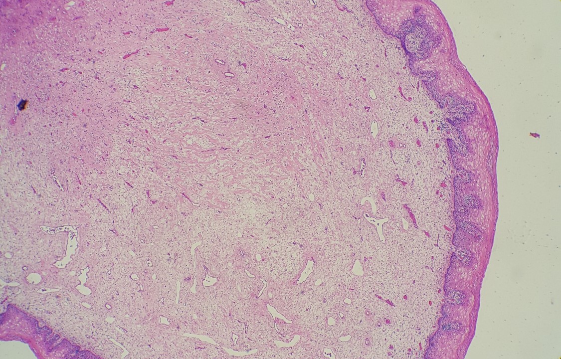
A male with an age of mid 60’s underwent cholecystectomy due to cholelithiasis. The patient’s preoperative CT scan (Fig. 1) and a microscopic image of the cholecystectomy specimen (Fig. 2) are below. What is your diagnosis and next thing to do?
- Chronic cholecysitis with cholelithiasis. No further study is required.
- Hyalinizing cholecysitits. Re-examination of the specimen with additional sections is required.
- Gallbladder adenocarcinoma. Staging is required.
- Improper section- additional sections and/or recut of the block with decalcification is(are) required.

Answer: B
It is a rare case of hyalinizing cholecystitis (HC) where the gallbladder (GB) wall calcification is highlighted on the CT scan image (Fig. 1 arrow). Microscopic findings (Fig. 2) show that the gallbladder wall is replaced by dense, hyalinized tissue transmurally. No epithelial layer or Rokitansky-Aschoff sinus is noted (Fig. 2 inset: inflammatory infiltrate only). Hence the anatomical landmarks (e. g., mucosa, muscularis) of the GB are no longer discernible.
“Porcelain GB” is a clinical/radiological diagnosis characterized by calcification of gallbladder wall, which has not been correlated with a pathologic diagnosis. Patel et al suggested that complete intramural calcification (complete porcelain GB) is an entity separated from HC which encompasses incomplete porcelain GB.
Hyalinizing cholecystitis and Porcelain GB are known to have an increased risk of developing GB adenocarcinoma. Patel et al and Stephen et al also revealed that hyalinizing cholecystitis with no or minimal calcification or selective mucosal calcification (incomplete porcelain) appears to be at higher risk of carcinoma. Due to the risk of malignancy, extensive sampling and meticulous microscopic examination are required once HC is identified.
References:
Patel S, Roa JC, Tapia O, Dursun N, Bagci P, Basturk O, Cakir A, Losada H, Sarmiento J, Adsay V. Hyalinizing cholecystitis and associated carcinomas: clinicopathologic analysis of a distinctive variant of cholecystitis with porcelain-like features and accompanying diagnostically challenging carcinomas. Am J Surg Pathol. 2011 Aug;35(8):1104-13.
Stephen AE, Berger DL. Carcinoma in the porcelain gallbladder: a relationship revisited. Surgery. 2001 Jun;129(6):699-703.
Case contributed by: Goo Lee, M.D.,Assistant Professor, Anatomic Pathology