Case History
76-year-old female with abdominal pain and rectosigmoid mass and omental nodules on CT scan. Resection of uterus, ovaries and sigmoid colon reveals 12.7 cm mass with tumor involving ovaries, uterus, and colon. Microscopic examination revealed a malignant tumor with two distinct growth patterns identified: one with solid tumor and second with glandular pattern.
Each tumor area showed different immunohistochemical result:
Solid tumor: Positive for CK7, PAX8; negative for CK20, CDX2
Glandular tumor: Positive for CK20, CDX2; negative for CK7, PAX8
Choose the correct diagnosis:
- Colon adenocarcinoma with undifferentiated tumor
- Carcinoma of mullerian origin with mucinous differentiation
- Collision tumor of ovarian and colon origin
- Mullerian adenocarcinoma, mixed epithelial type
Answer: C. Collision tumor of ovarian and colon origin
Explanation:
Collision tumors are rare neoplasms which consist of two or more distinct neoplasms that develop adjacent to one another and coexist with no or minimal intermingling between them.
We present a rare collision tumor composed of papillary serous adenocarcinoma of Mullerian origin involving uterus and bilateral ovaries, and a mucinous adenocarcinoma of the sigmoid colon.
Resection of the uterus, ovaries, and sigmoid colon revealed a 12.7 cm in combined maximal dimension. Microscopically malignant tumor with two distinct growth patterns was identified: one with solid tumor and second with glandular pattern.
The uterus and ovaries were diffusely involved by solid papillary serous adenocarcinoma (CK7 and PAX8 positive; CK20 and CDX2 negative immunohistochemical stains). Colon showed invasive mucinous adenocarcinoma with an opposite staining pattern.
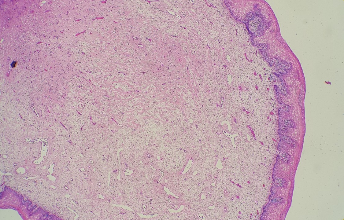
Collision of both tumors is demonstrated in Figure A (H&E stain). Figure B shows positive PAX8 stain limited to papillary serous adenocarcinoma (lower left). Figure C demonstrates positive CK20 stain only in colonic adenocarcinoma (upper right).
Recognition of collision tumors is clinically important. Their diagnosis is often incidental and their behavior remains widely unknown.
References:
Collision Tumors of the Gastrointestinal Tract: A Systematic Review of the Literature. Schizas D, Katsaros I, Michalinos A, Damaskos C, Garmpis N, Ntomi V, Agrogiannis G, Stergiopoulos S, Tsaroucha AK., Anticancer Res. 2018 Nov;38(11):6047-6057. doi: 10.21873/anticanres.12955. PMID: 30396919
Case contributed by: Lea Novak, Associate Professor, Anatomic Pathology