Case History
The patient is a 45-year-old HIV+ male with hematemesis and weight loss. Endoscopy reveals maroon-red distal esophageal plaques and nodules.
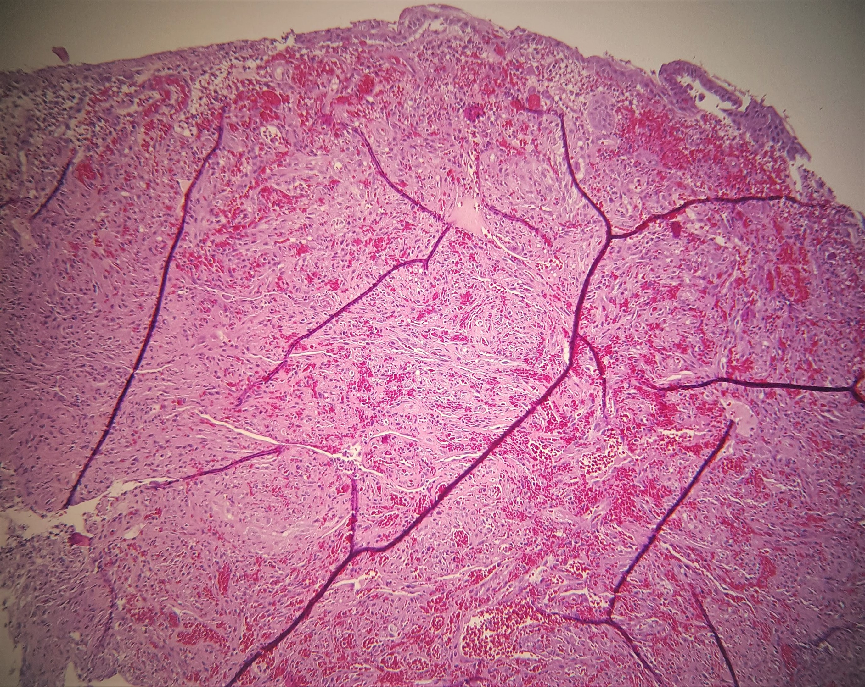
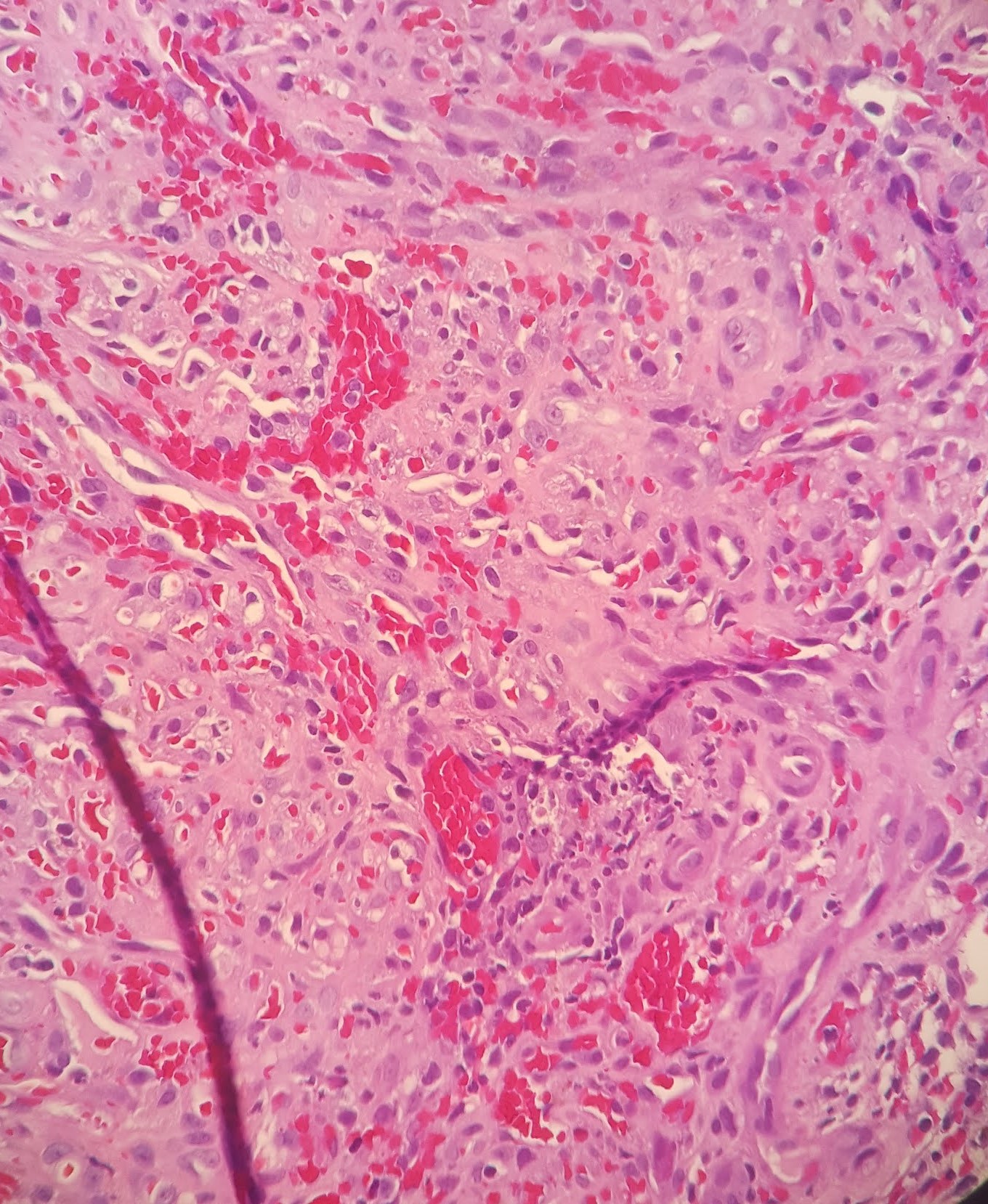
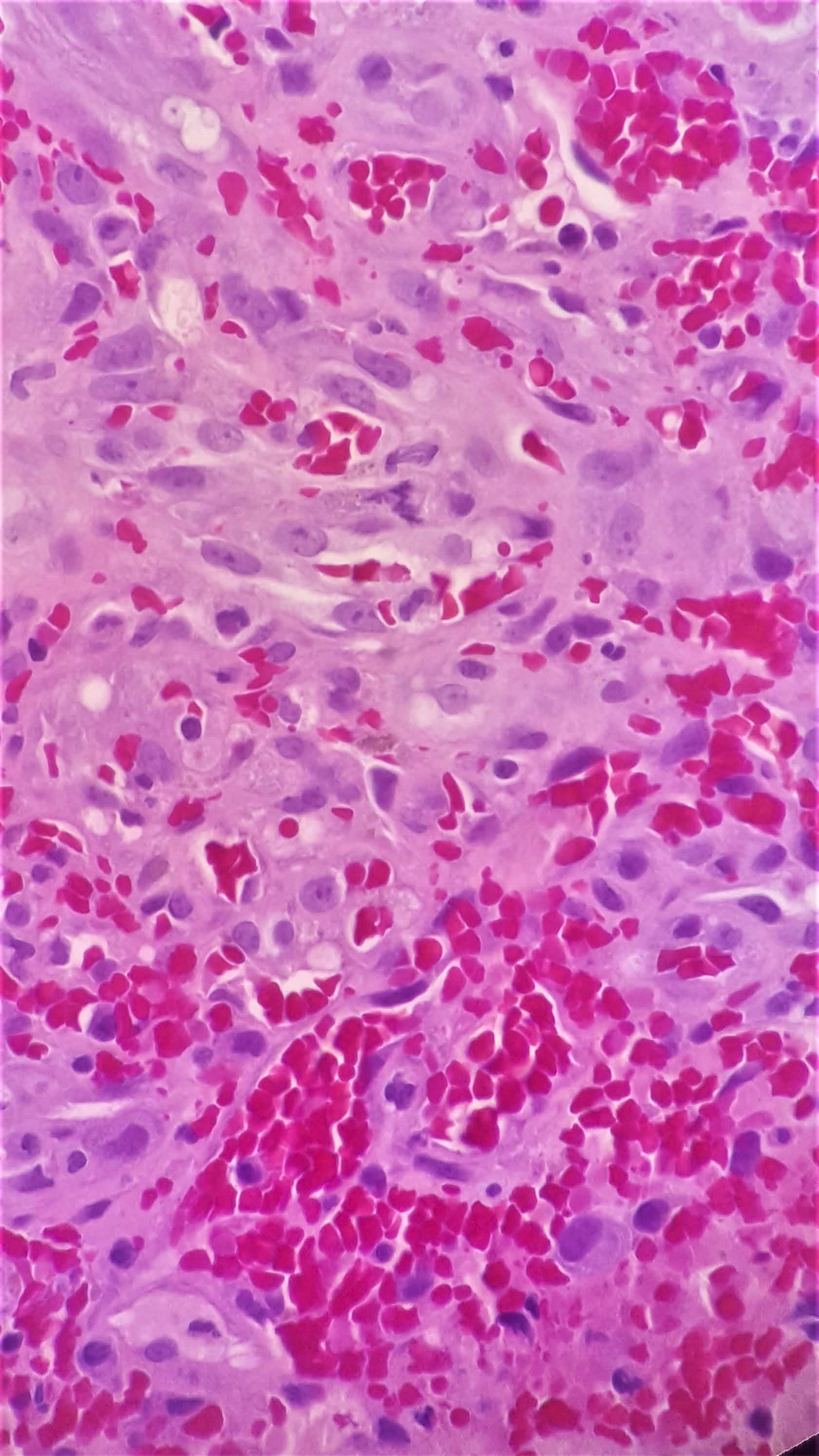
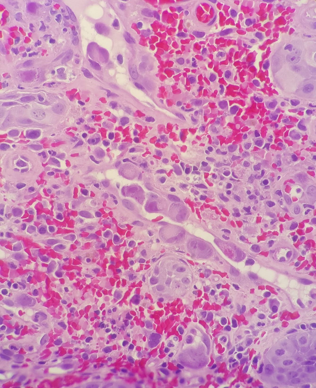
Microscopic examination reveals the following.
What is your diagnosis of this case?
- CMV Esophagitis
- Angiosarcoma
- Kaposi Sarcoma + HSV Esophagitis
- Kaposi Sarcoma + CMV Esophagitis
Correct Answer: D, Kaposi Sarcoma + CMV Esophagitis
Discussion:
Immunohistochemistry for HHV8 and CMV were both positive on this case, confirming the diagnosis as Kaposi sarcoma with superimposed CMV esophagitis.
Kaposi sarcoma is the most common GI malignancy in the setting of AIDS disease. Gastrointestinal Kaposi sarcoma are commonly asymptomatic but may show clinical symptoms of hemorrhage, nausea, weight loss, etc.
Histologically, Kaposi sarcoma is composed of monomorphic spindle cell proliferation arranged in a vague fascicular appearance with intervening slit-like spaces. Intracellular and extracellular eosinophilic PAS+ (diastase-resistant) hyaline globules are present as well as extravasated red blood cells. HHV8 (LANA) immunohistochemical stain can confirm the diagnosis. Treatment is variable based on the extent of the patient’s disease.
CMV esophagitis is the second most common GI manifestation of cytomegalovirus infection after colitis. CMV infections are common in immunocompromised patients but may be seen in immunocompetent patients in rare cases. Endoscopically, CMV esophagitis shows a non-specific appearance of inflammatory exudate and ulceration.
Histologically, CMV infected cells show enlarged cells (cytomegaly) and enlarged nuclei with ovoid intranuclear inclusions with a surrounding clear halo. This is classically called the “Owl’s Eye” appearance. Immunohistochemistry for CMV can confirm the presence of CMV infected cells. CMV infections can be treated with antiviral therapies and is important to notify the treating physician as early as possible.
Reference:
Arora, M., & Goldberg, E. M. (2010). Kaposi sarcoma involving the gastrointestinal tract. Gastroenterology & hepatology, 6(7), 459–462.
Case contributed by: Chirag Patel, M.D., Assistant Professor, Anatomic Pathology