Case History
A 50-year-old presents with multiple liver masses. Histologic examination reveals the following:
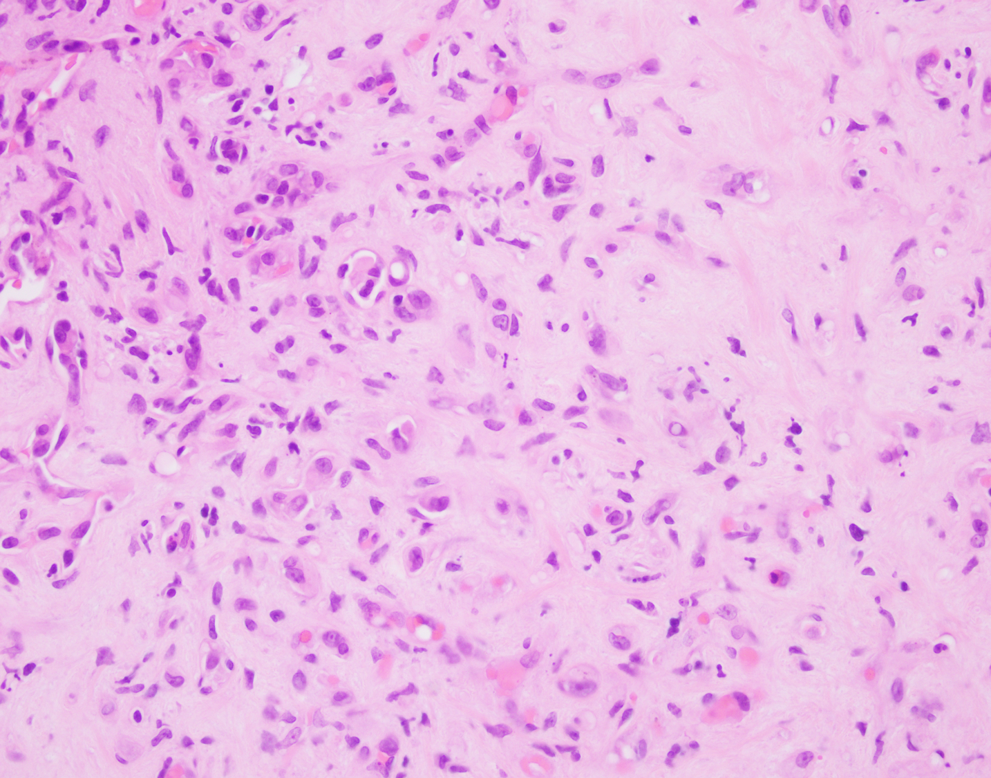
Figure 1: Hyalanized stroma with scattered cells
Figure 2: Haphazardly arranged atypical cells within the stroma, some showing intracytoplasmic vacuole/lumina
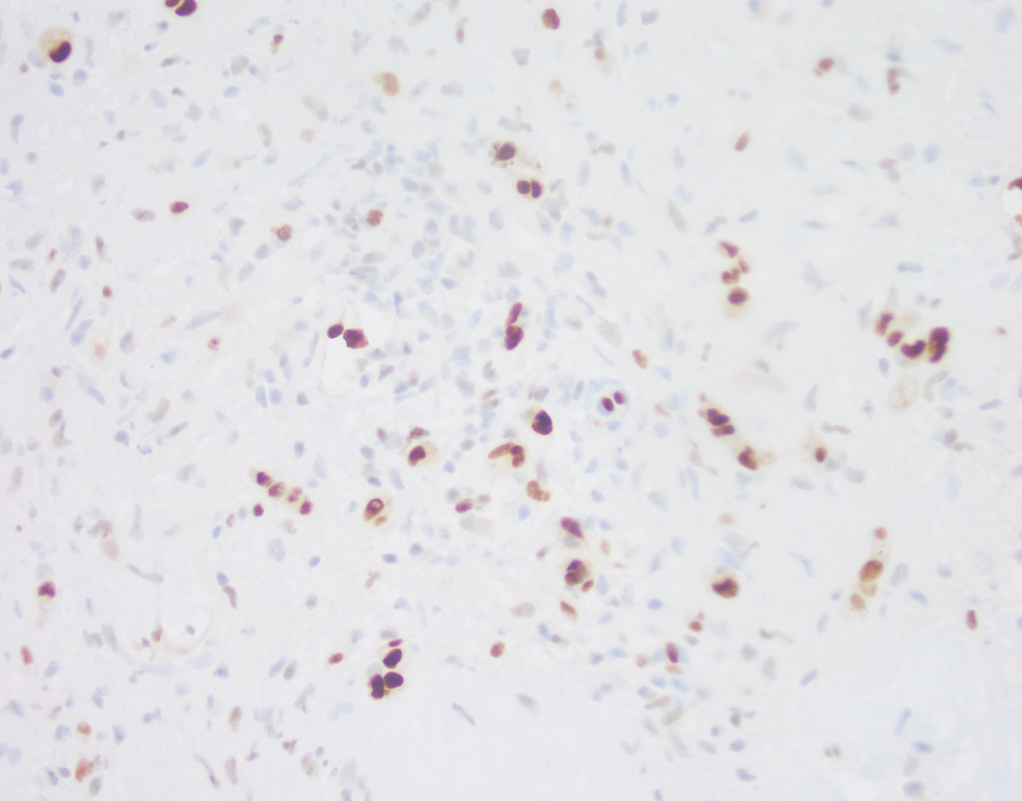
Figure 3: ERG immunostain highlights those atypical cells

What is the diagnosis?
- Epithelioid hemangioma
- Epithelioid hemangioendothelioma
- Angiosarcoma
- Inflammatory pseudotumor
The answer is: "B" Epithelioid Hemangioendothelioma.
Explanation:
This tumor typically shows scattered mildly atypical cells in a background of fibromyxoid stroma. The diagnostic feature is the presence of intracytoplasmic vacuole/lumina formation. The atypical epithelioid cells are positive for ERG and CD31, supporting the vascular nature of this tumor. The differential diagnosis includes carcinoma, particularly metastatic, and angiosarcoma. Lack of marked atypia argues against the diagnosis of angiosarcoma. The keratin stain was negative in this case, which along with the positivity for vascular markers, rules out the possibility of carcinoma, though keratin positivity can be rarely seen in epithelioid vascular tumors.
Epithelioid hemangioendothelioma is a rare low-grade vascular malignancy that occurs in adults. The tumor is usually multifocal. It typically grows around pre-existing structures. Entrapped portal tract/bile duct can be seen in the tumor, as seen in this case.
Reference: Studer LL, Selby DM. Hepatic Epithelioid hemangioendothelioma. Arch Pathol Lab Medicine. 2018;142:263-267
Case contributed by: Deepti Dhall, M.D., Professor, Anatomic Pathology