Case History
A 21-year-old female presented with anemia and thrombocytopenia. MRI of the abdomen performed demonstrated a rounded well-defined 5.9 × 7.8 × 8.1 cm anterior subcapsular mass present within the spleen. Laparoscopic hand-assisted splenectomy was performed.
What is the best diagnosis?
- Hamartoma of spleen
- Angiosarcoma
- Sclerosing angiomatoid nudular transformation of the spleen (SANT)
- Littoral cell angioma
Answer:
C. Sclerosing angiomatoid nudular transformation of the spleen (SANT)
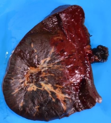
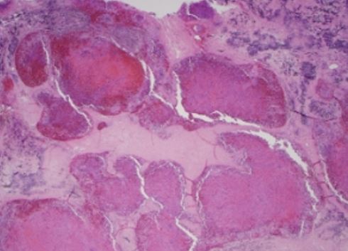
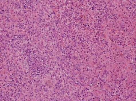
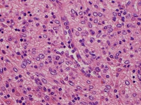
Pathologic analysis reported a 445 g spleen measuring 14.5 × 10 × 7.5 cm with a 7.5 × 7 × 5.5 cm well-circumscribed nonencapsulated dark red mass with tan-yellow to tan-white fibrous tissue expanding radially from the center of the lesion (Figure A). Histopathologic and immunohistochemical evaluation of the splenic mass revealed the mass was composed of thick dense fibrous bands which coalesced to form angiomatous nodules composed of a fine network of numerous blood vessels surrounded by collagenous stroma (Figure B). An increased number of plasma cells, rare small lymphocytes, rare neutrophils, and no eosinophils were seen within the vascular nodules (Figure C). A population of bland spindle and/or plump endothelial cells with large oval nuclei, vesicular chromatin, and prominent nucleoli was noted (Figure D). The dense fibrous bands contained numerous hemosiderin-laden macrophages and focal areas of microcalcifications. Numerous arteries of variable size with thickened hyalinized walls. These vessels stained positive for CD31, CD8, CD34, Factor VIII, and rare focal D240. CD68 highlighted an increased number of Littoral cells/histiocytes. The angiomatous nodules contained cells positive for CD31, CD8, CD34, vimentin, SMA, focal positive for EMA, rare positive S100, and negative for desmin. Approximately 10% positivity for Ki-67. Additional CD21 stains highlighted rare positive cells. The histopathologic findings were consistent with sclerosing angiomatoid nodular transformation (SANT).
Sclerosing Angiomatoid Nodular Transformation (SANT) is a benign vascular lesion of the spleen. The differential diagnosis includes hemangioma and littoral cell angioma, both lack the distinctive histological features of SANT.
Reference:
Gooch C., Gaballah, A, Nelson J, Wade M, Chen R, Sclerosing angiomatoid nodular transformation of the spleen: a case report of thrombocytopenia and a hypervascular splenic mass, Radiol Case Rep. 2019 Apr; 14(4): 521-525
Case contributed by: Rong Chen, M.D., Surgical Pathology Fellow, UAB Pathology