Case History
A 79-year-old Caucasian male was diagnosed with CLL in 8/2011. His CT revealed splenomegaly and some borderline enlarged nodes. One in the left axilla was 1.4cm. In January, 2017, his CT showed mild progression of his nodes. The largest one was left external iliac lymph node 3.4 x 1.2 cm, with a 1.4 cm increase over 13 months. In March 2019, he developed cough, chest X-ray reveal bronchial changes. In several month, he also developed peritoneal fluid.
What is the diagnosis?
A: Reactive mesothelial cells
B: Lymphoma
C: Melanoma
D: Mesothelioma
Answer:
"C" Melanoma
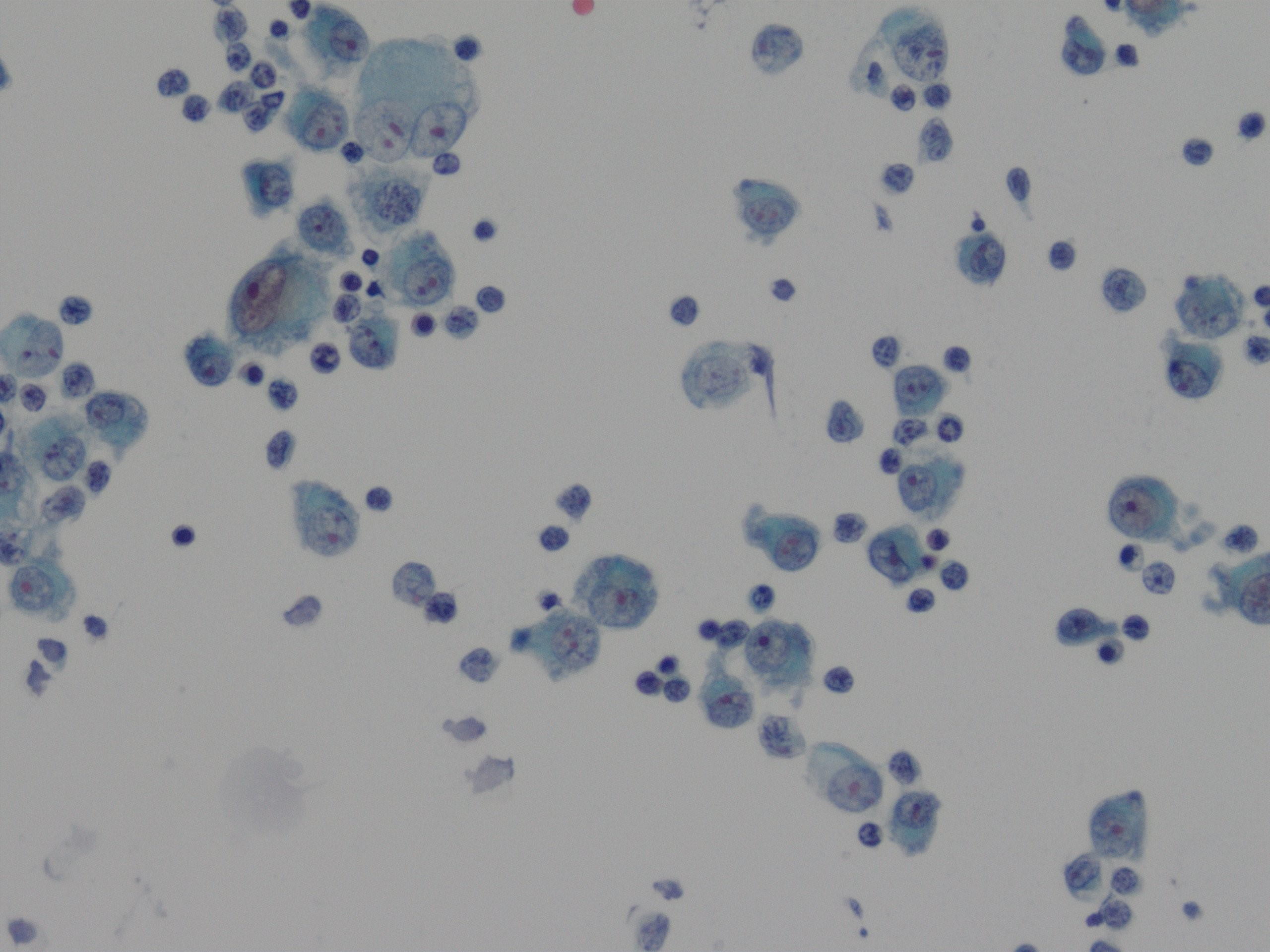
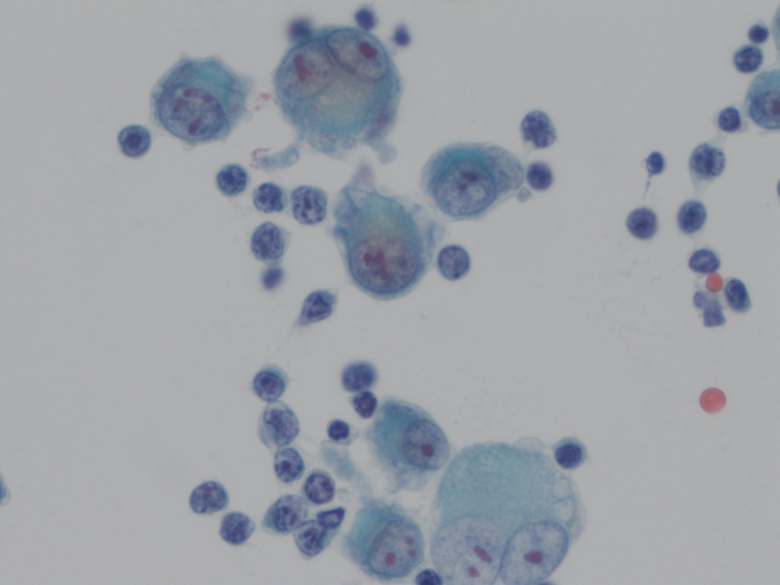
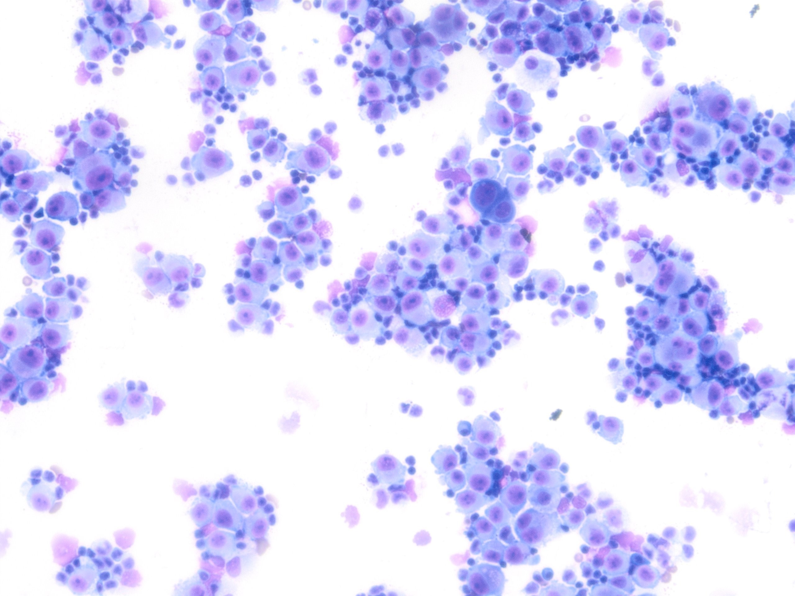
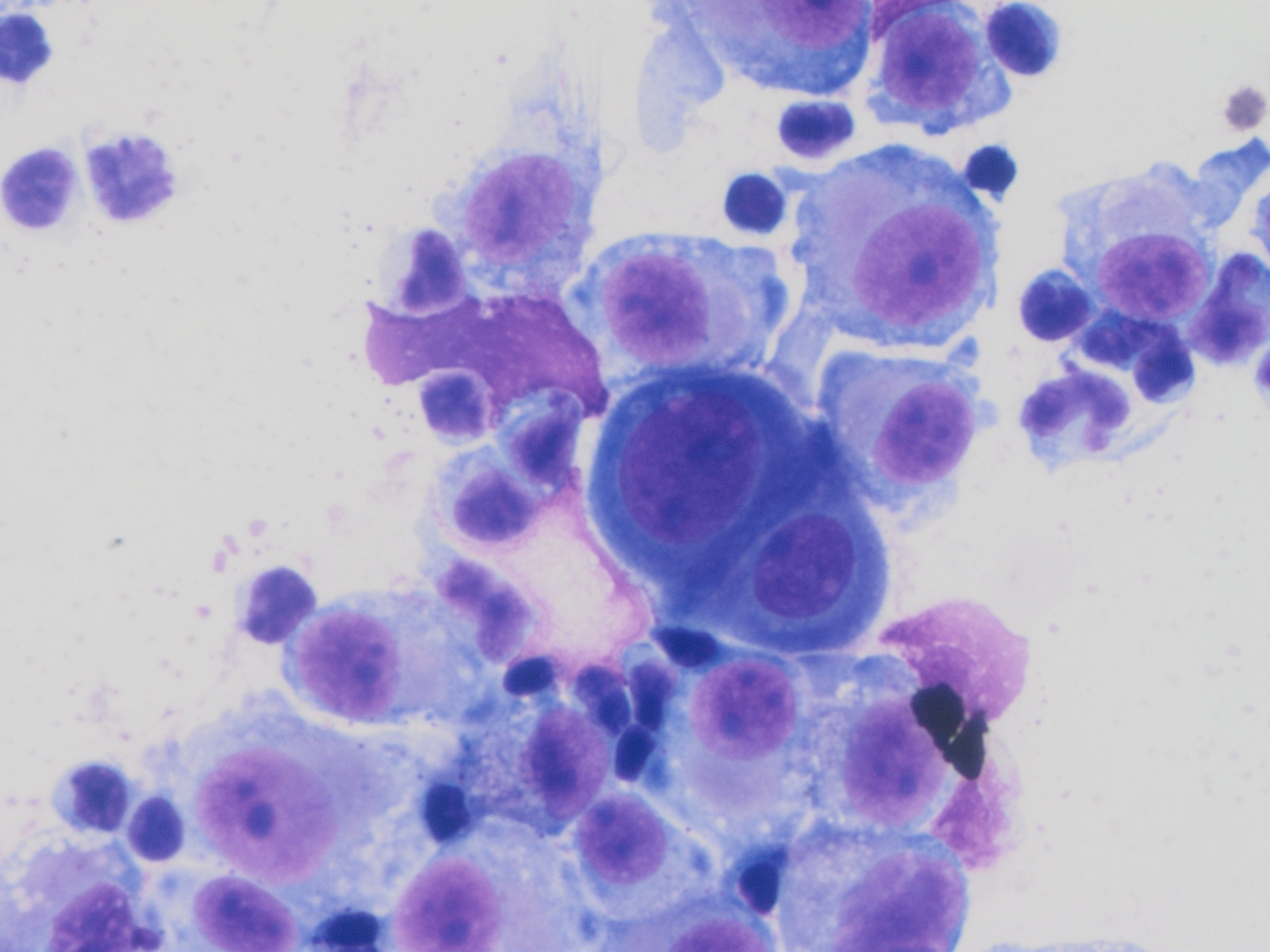
The diagnosis of melanoma can be subtle in effusions. The malignant cells resemble mesothelial cells. They are usually isolated and loosely clustered. In some cases, cells show a fine brown cytoplasmic pigmentation. If malignant cells are predominant, there would be no "second" population. A clue of this case is that more than 90% percent of the malignant cells are plasmacytoid, which raises the concern for melanoma. Reactive mesothelial cells could have plasmacytoid feature with prominent nucleoli, but they would not be uniformly plasmacytoid. In this case, Immunostain shows the malignant cells are positive for SOX-10.
Case contributed by: Shoujun Chen, M.D., Ph.D., UAB Cytopathology Fellow