Case History
A 74 -year-old woman with a clinical history of dementia dies at home. The family wishes to clarify the history of dementia. An autopsy shows cortical atrophy and ventriculomegaly. Based on the 3 images taken from sections of frontal lobe, what is the most likely diagnosis?
- Dementia with Lewy Bodies
- Vascular dementia
- Alzheimer dementia
- Frontotemporal dementia
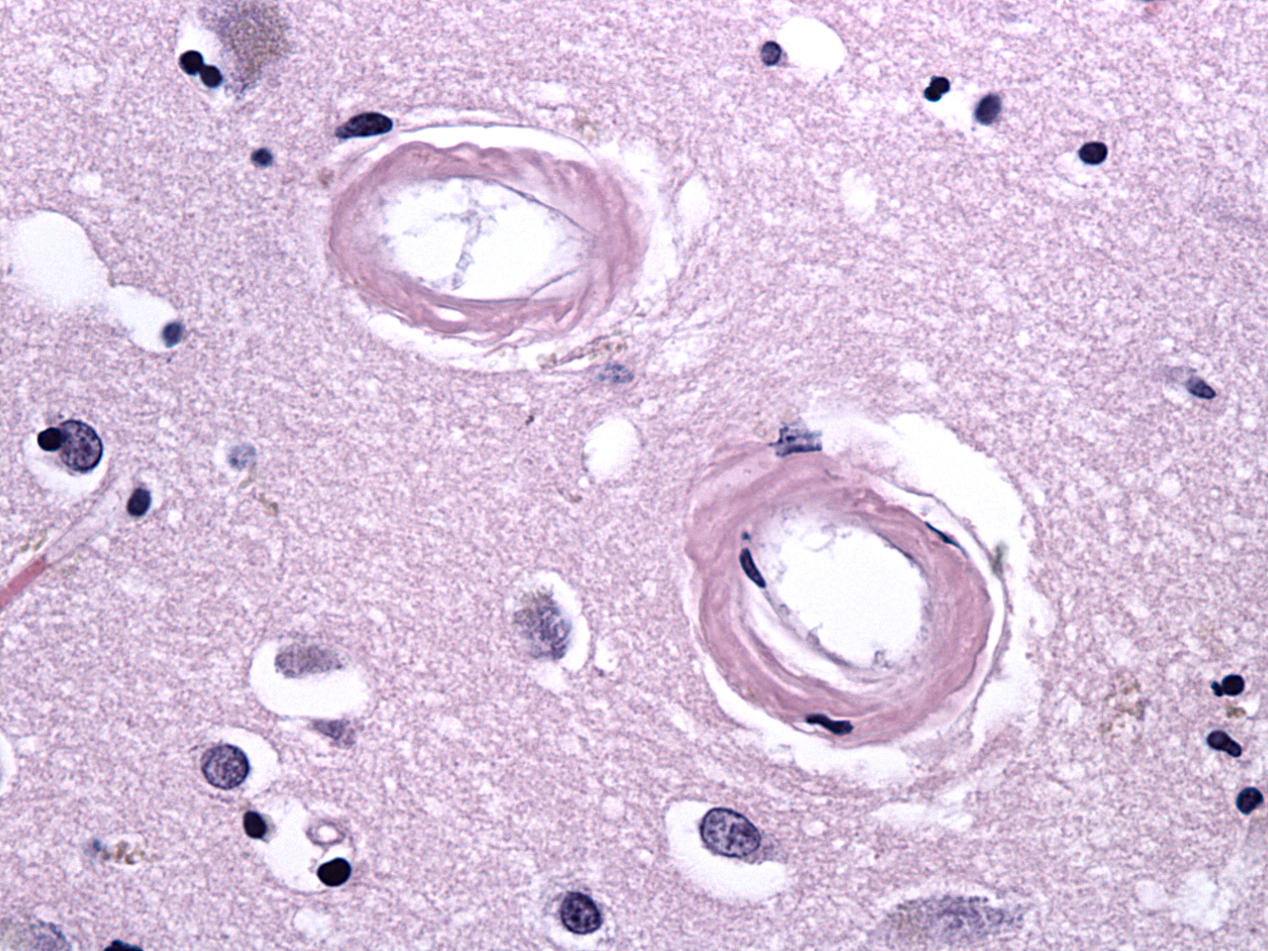
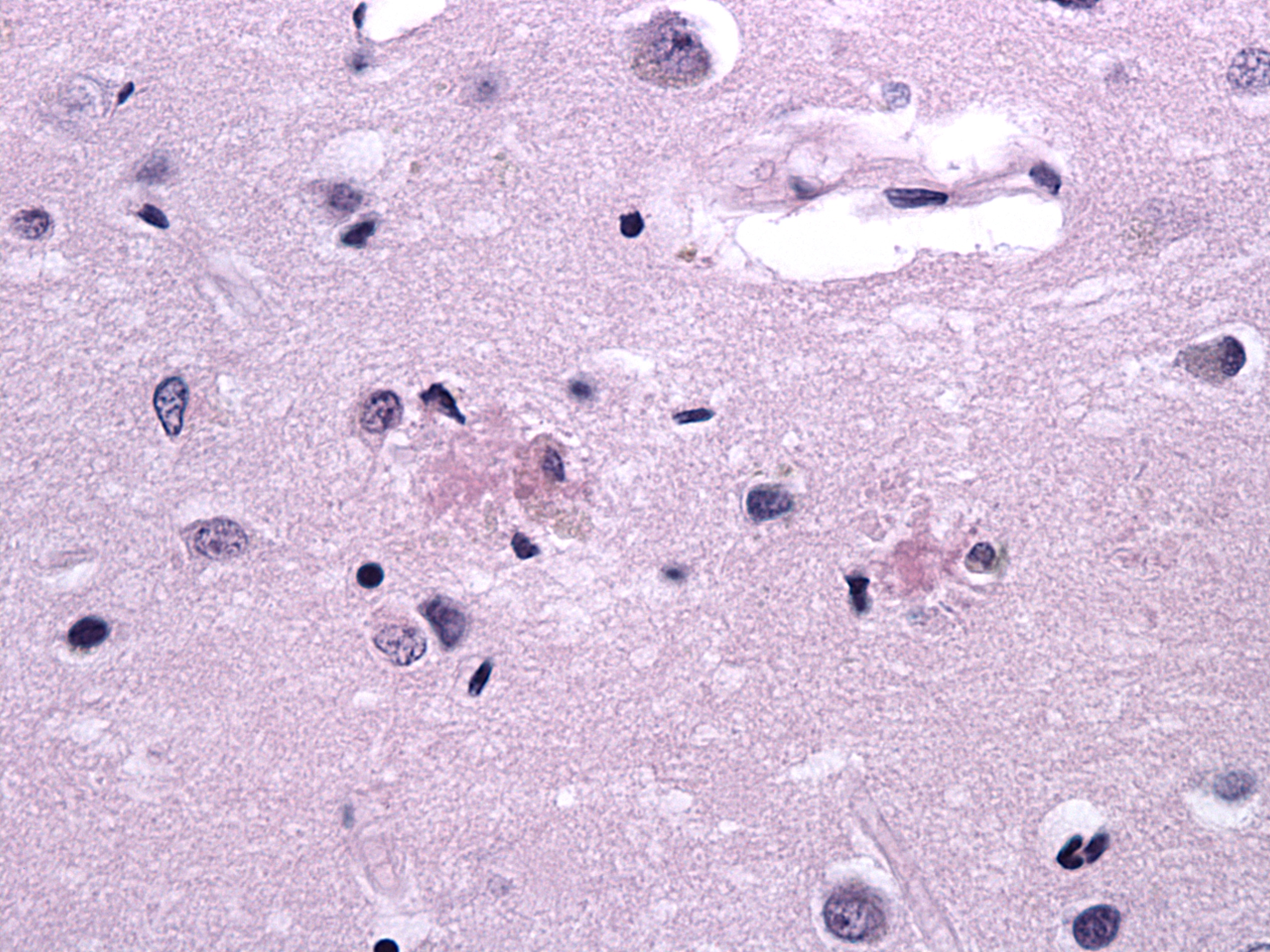
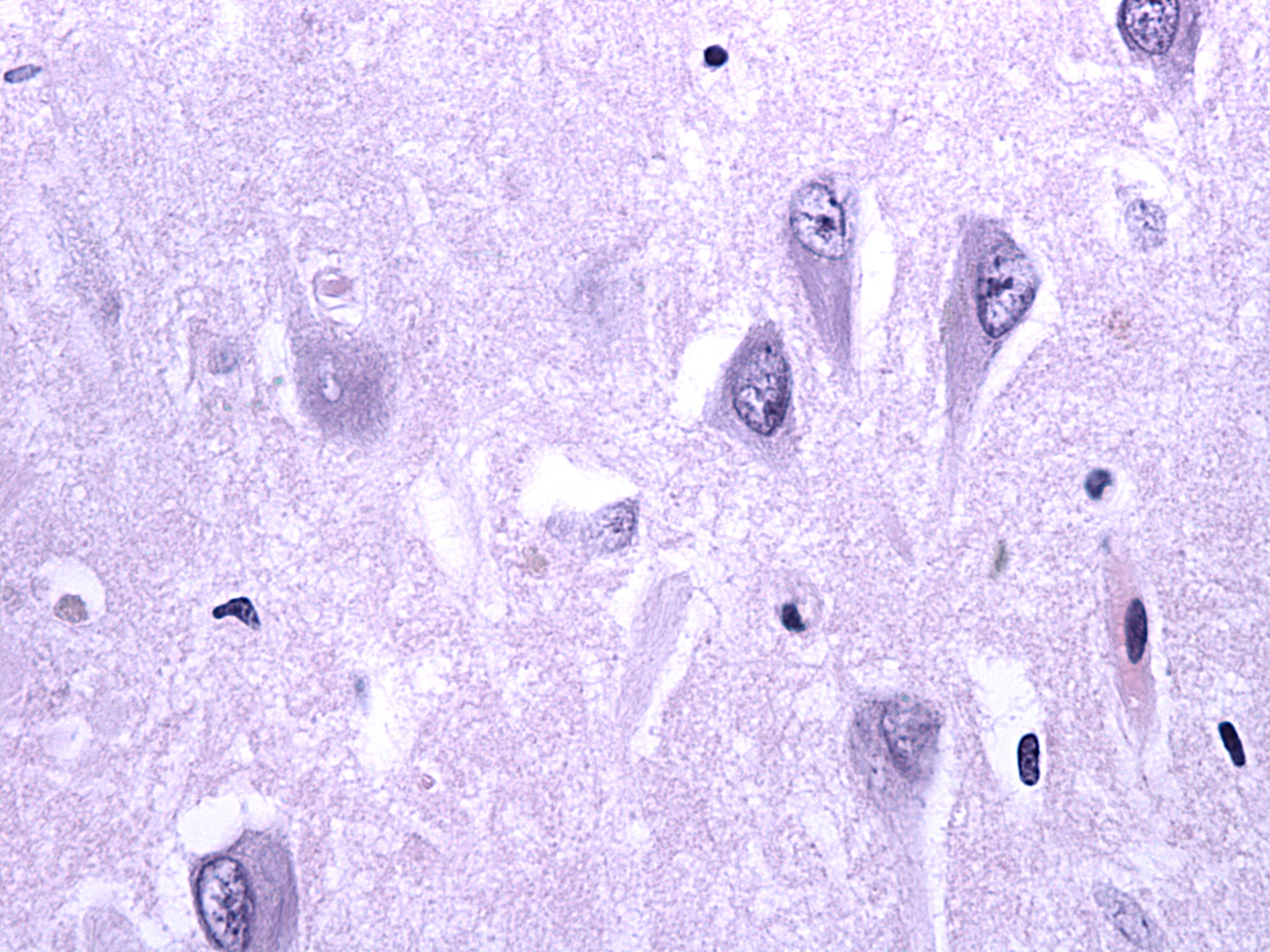
Answer: C: Alzheimer's disease.
Discussion: A critical feature is an adequate density of neuritic plaques and neurofibrillary tangles in neocortex. The presence of vessels suspicious for cerebral amyloid angiopathy is also helpful. If plaques and tangles are limited to the hippocampus, they could simply represent age-related changes. This case also illustrates that many such findings can be seen on H&E with careful observation. Of course, we always confirm with appropriate stains!
Case contributed by: James Hackney, M.D., Associate Professor, Neuropathology, and Astin Powers, M.D., UAB Neuropathology Fellow