Case History
A 12-year-old boy presented with a lytic lesion in the epiphysis of the proximal tibia with sclerosis at the periphery on imaging. A biopsy was performed.
- Giant cell tumor of bone
- Solid variant of aneurysmal bone cyst
- Giant cell-rich osteosarcoma
- Chondroblastoma
The answer is “D”, Chondroblastoma
Chondroblastoma is a relatively rare lesion, representing approximately 1% of bone tumors. It usually arises in skeletally immature patients, mostly affecting bones around the knee, followed by the proximal humerus. Rare tumors arise in the temporal bone or calcaneus. Patients typically present with joint pain, swelling, and decreased range of motion. Radiologically, chondroblastoma is sharply delineated, lytic epiphyseal lesion rimmed by sclerotic bone. Large lesions may cross the growth plate and involve the metaphysis.
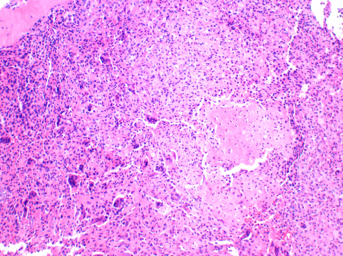
Histologically, chondroblastoma shows ovoid-to-round mononuclear chondroblasts with characteristic nuclear grooves, eosinophilic cytoplasm, and well defined cell borders. Variably sized eosinophilic to amphophilic fibro-chondroid islands are commonly seen. Approximately one-third of cases show fine “chicken-wire” calcifications surrounding individual tumor cells. Also present are dispersed osteoclast-type multinucleated giant cells. Secondary aneurysmal bone cyst formation is not uncommon. True hyaline cartilage is almost never seen in chondroblastoma.
H3F3B driver mutations characteristically define chondroblastoma. This is in contrast to H3F3A mutations typically seen in giant cell tumor of bone, another giant cell-rich tumor that affects epiphysis of long bones in the skeletally mature individuals.
Most chondroblastomas of long bones are successfully treated by curettage, with the remainder showing local recurrence. Temporal bone and calcaneus lesions tend to show higher rates of recurrence. Malignant transformation is exceedingly rare.
References
- Wei S, Siegal GP, eds. Atlas of Bone Pathology. New York. Springer. 2013:122-126.
- Behjati S, et al. Distinct H3F3A and H3F3B driver mutations define chondroblastoma and giant cell tumor of bone. Nat Genet. 2013 Dec;45(12):1479-82.
Case contributed by: Shi Wei, M.D., Ph.D., Professor, Anatomic Pathology, UAB Pathology