Case History
The patient, a 57-year-old female, was found to have 15.5 cm unilateral right adnexal mass. No evidence of extraovarian/peritoneal involvement was seen intraoperatively. The right ovarian tumor consisted of a multiloculated cyst with smooth inner lining. There were no ovarian surface involvement identified grossly. Representative microscopic images are shown. Immunohistochemical studies are pending. Based on the H&E appearance, the most likely diagnosis is:
- Mucinous cystadenoma
- Mucinous borderline tumor
- Seromucinous borderline tumor
- Secondary ovarian involvement by mucinous tumor of lower gastrointestinal tract origin

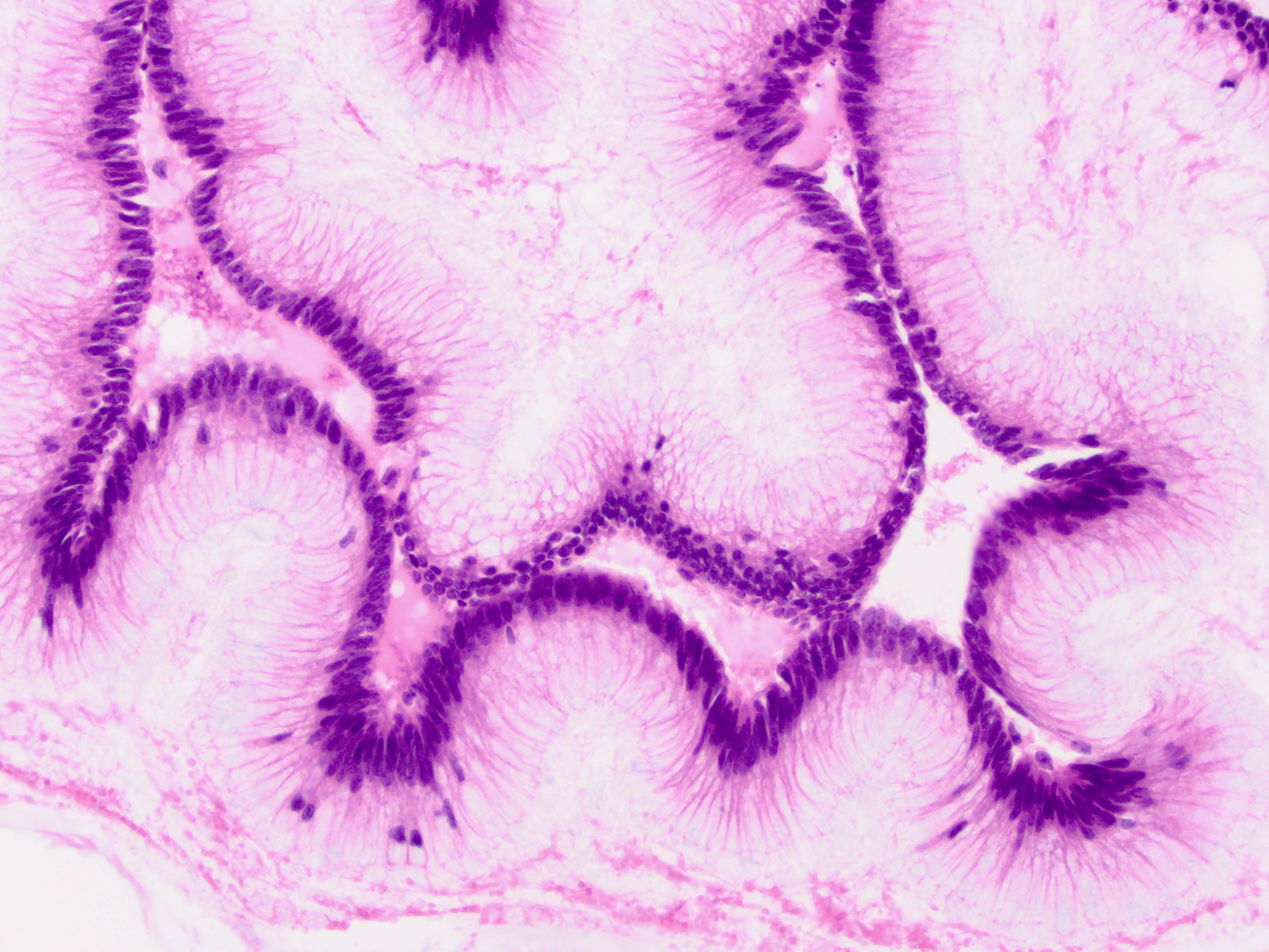
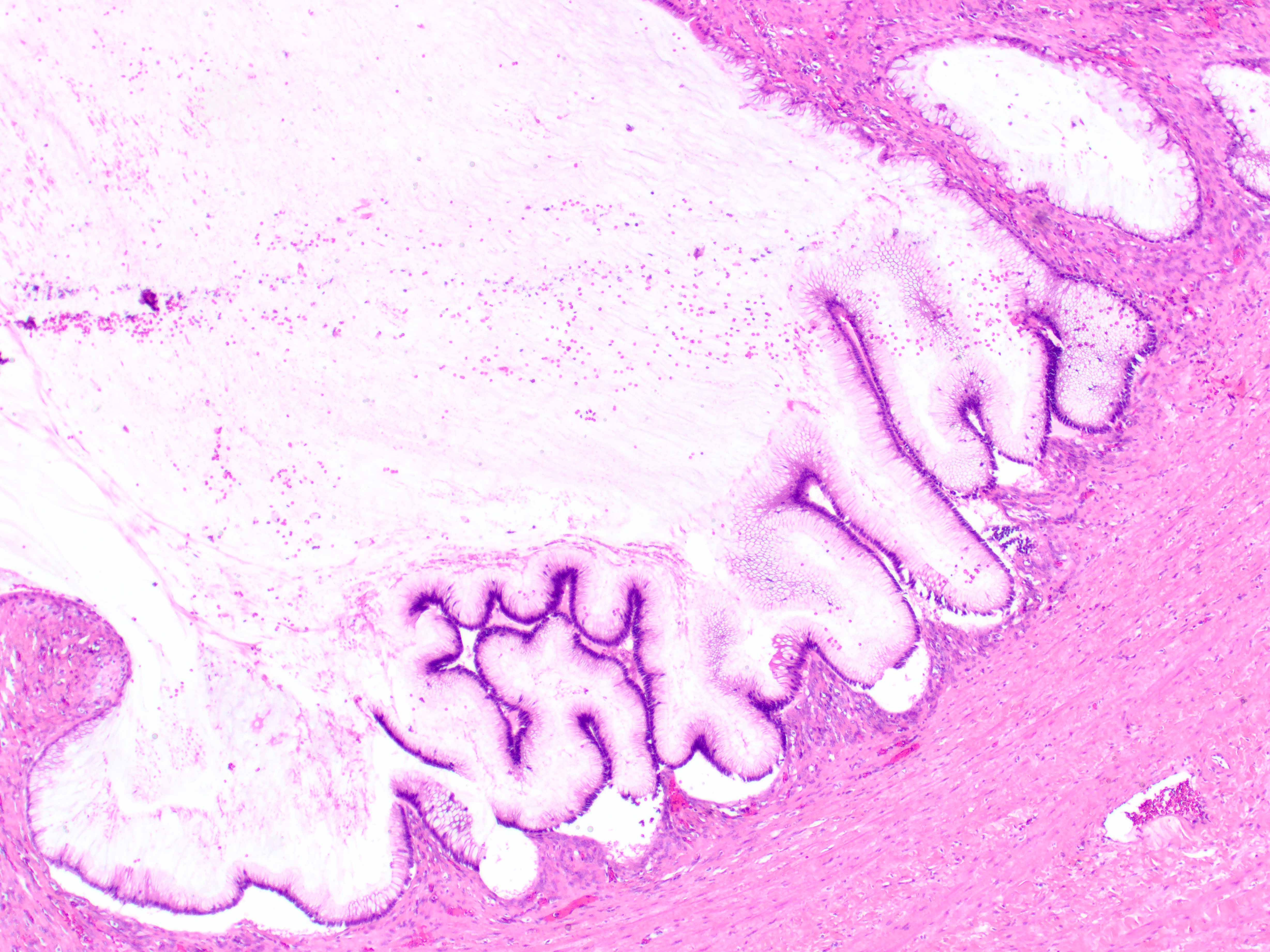
Correct answer: D--Secondary ovarian involvement by mucinous tumor of lower gastrointestinal tract origin.
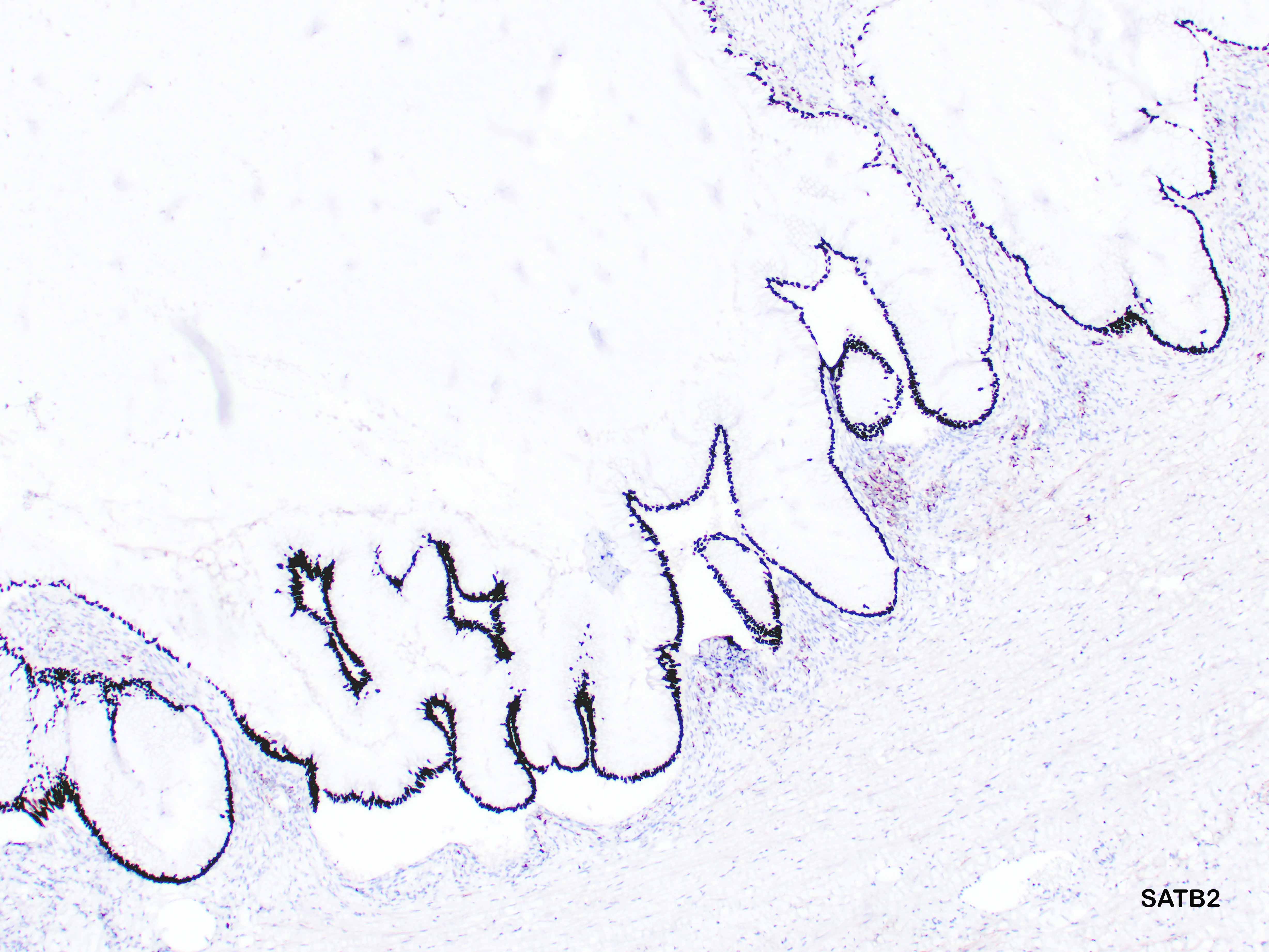
Immunohistochemical stains demonstrated that the tumor cells are diffusely positive for SATB2 (see figure) and CK20 and negative for CK7, PAX8, ER, and PR (not shown). While a large unilateral ovarian mucinous neoplasm is generally associated with an ovarian primary, this immunoprofile is not typical of that and strongly suggest secondary ovarian involvement by a neoplasm of lower gastrointestinal tract origin. The possibility of a mucinous neoplasm arising in a mature cystic teratoma has been considered, but no evidence of teratomatous elements was identified upon extensive sampling. Given bland/ low-grade appearance of lesional epithelium, an appendiceal low-grade appendiceal mucinous neoplasm should be considered. Appendix has not been sampled during this surgery. Therefore, additional clinical evaluation is required.
Case contributed by: Anna Yemelyanova, M.D., Hazel M. Gore Endowed Professor of Gynecologic Pathology