Case History
A 58-year-old male with a liver and omental mass. Omental mass biopsy shows:
- Signet ring cell carcinoma
- Ex Goblet cell carcinoid/Goblet cell adenocarcinoma
- Epithelioid Hemangioendothelioma
- Angiosarcoma
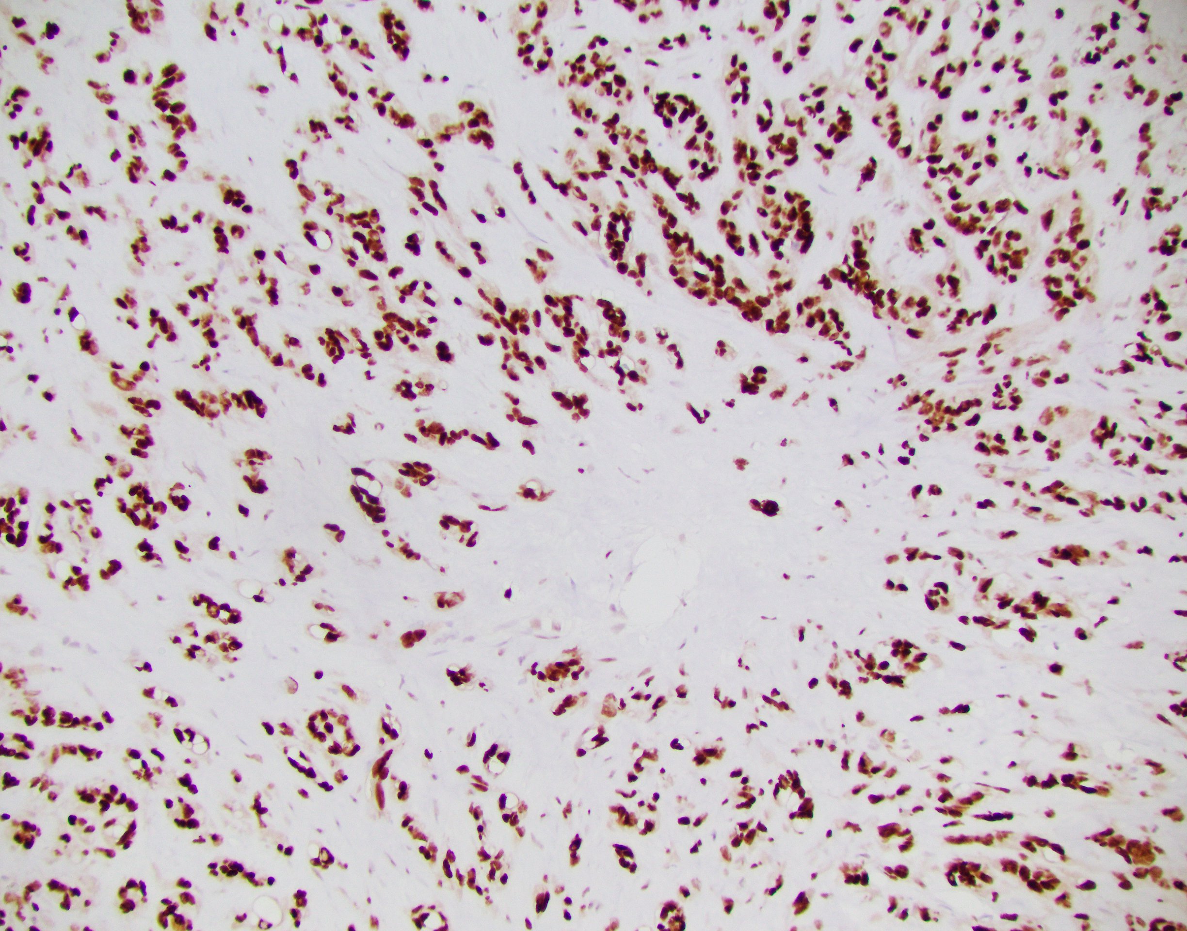 EH ERG2
 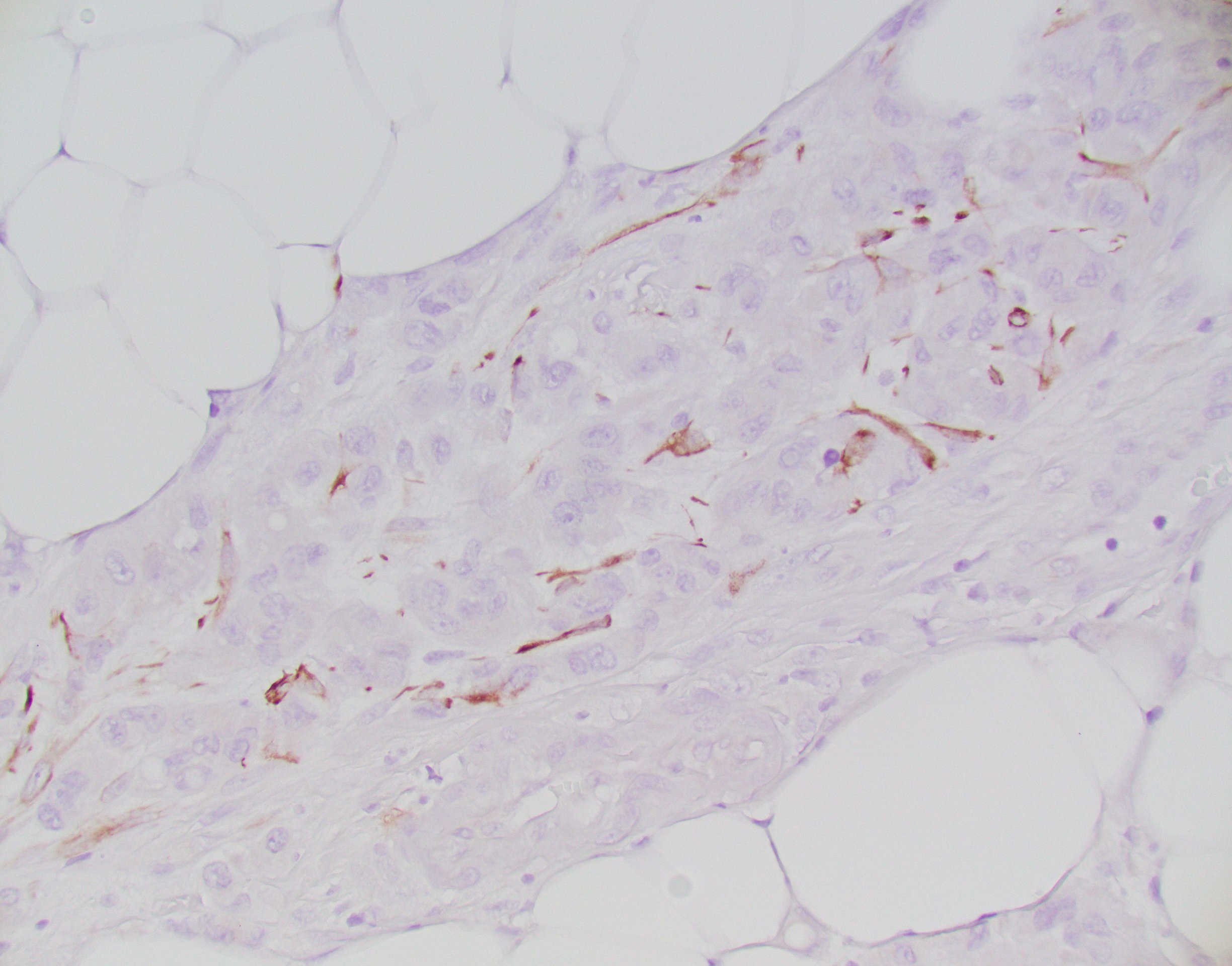 EH Keratin
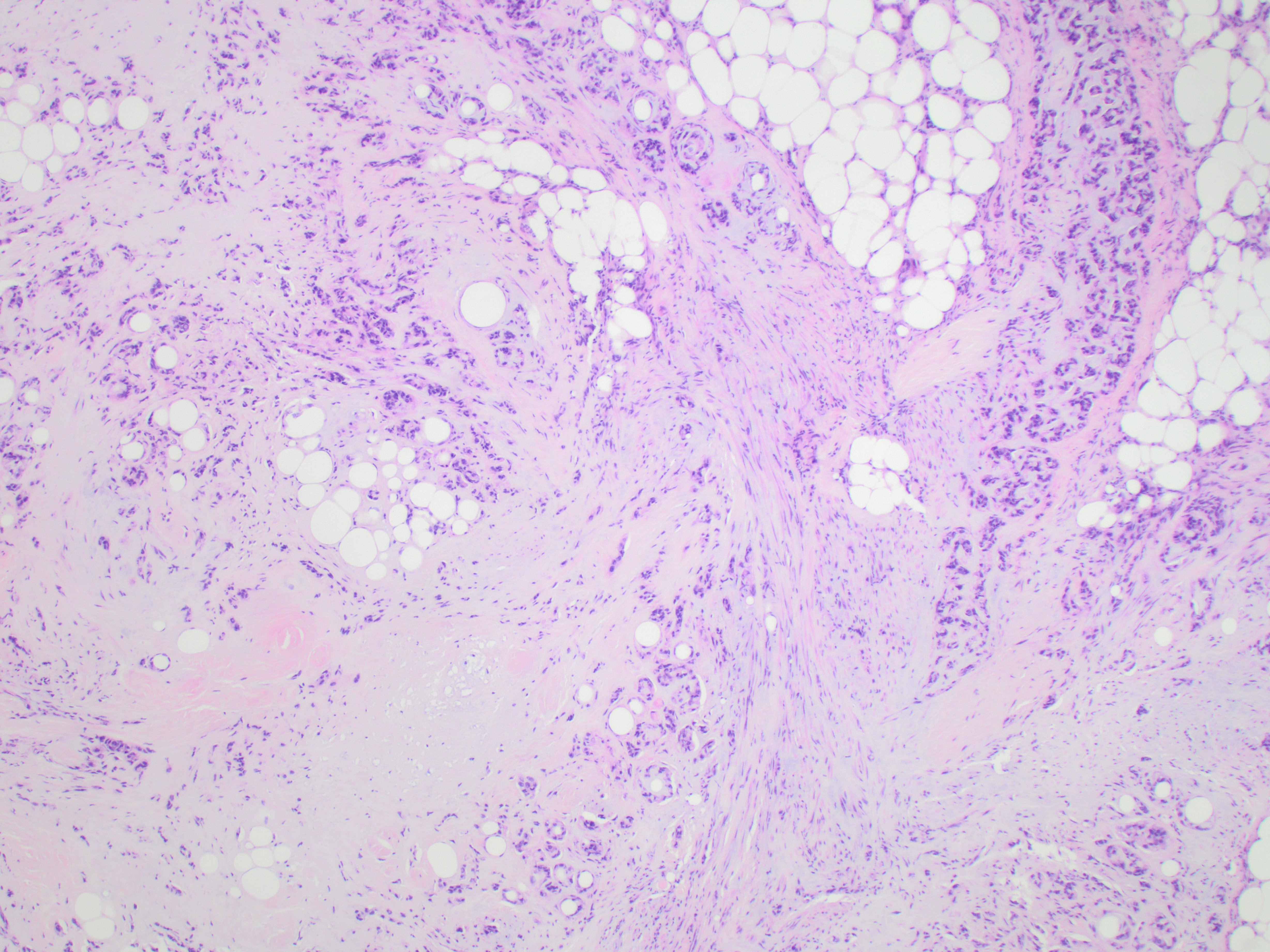 EH omentum 4x
 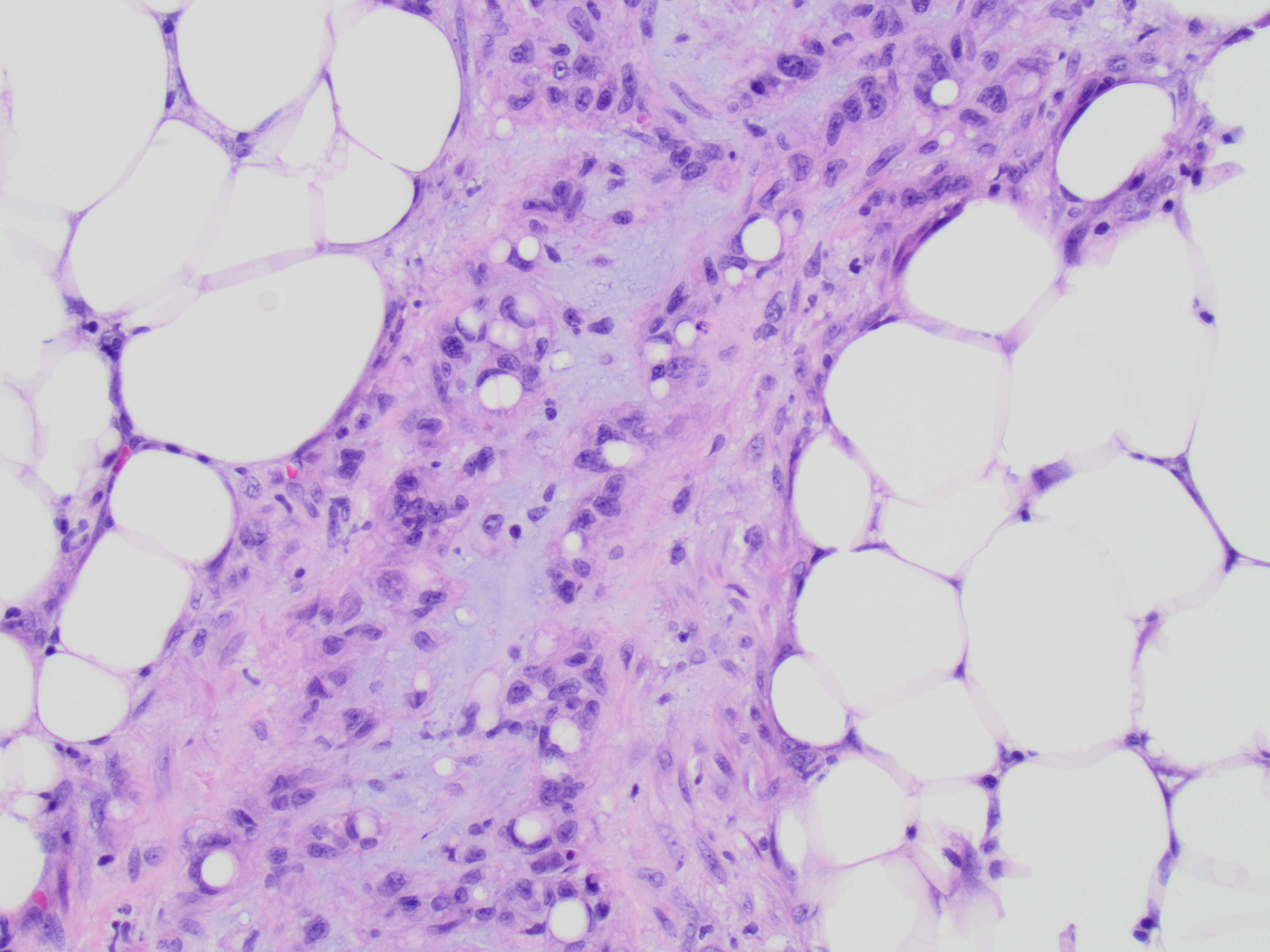 EH omentum 20x
Answer: C
Epithelioid Hemangioendothelioma is a malignant endothelial neoplasm composed of epithelioid cells in a myxohyaline or fibrous stroma. These may arise in the liver, lungs, bone, or soft tissue, often presenting with multifocal disease. By immunohistochemistry, the tumor cells are positive for endothelial markers (CD31, CD34, ERG) and may be potentially positive for keratins, leading to possible confusion with carcinoma. WWTR1-CAMTA1 gene fusion t(1;3)(p36;q25) translocation is a features of this lesion, found in 90% of cases.
Epithelioid Hemangioendothelioma is a malignant endothelial neoplasm composed of epithelioid cells in a myxohyaline or fibrous stroma. These may arise in the liver, lungs, bone, or soft tissue, often presenting with multifocal disease. By immunohistochemistry, the tumor cells are positive for endothelial markers (CD31, CD34, ERG) and may be potentially positive for keratins, leading to possible confusion with carcinoma. WWTR1-CAMTA1 gene fusion t(1;3)(p36;q25) translocation is a features of this lesion, found in 90% of cases.
Case contributed by: Chirag Patel, Assistant Professor, Anatomic Pathology