Case History
This is an 18-year-old female with bilateral adnexal masses.
What is the diagnosis?
- Granulosa cell tumor
- Sertoli Leydig cell tumor
- Dydgerminoma
- Lymphoma
Correct answer: C.
This patient presented with androgen insensitivity syndrome (AIS, formerly known as testicular feminization syndrome). She was referred to a gynecologist after report of no onset of menses by the age of 16 and was found to have 46XY karyotype and no uterus on imaging. AIS is an X-linked recessive condition resulting in a failure of normal masculinization of the external genitalia in chromosomally male individuals. Dysgenetic gonads are known to undergo malignant transformation. Therefore, bilateral gonadectomy is performed.
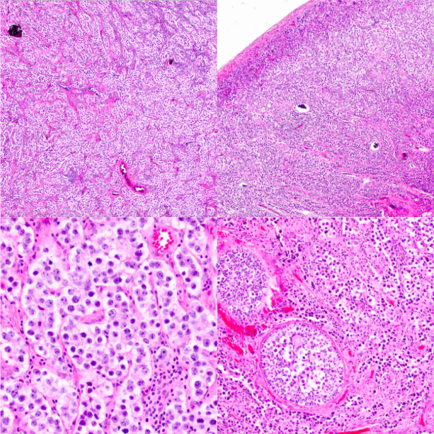
The tumor is composed of relatively uniform large cell with vesicular chromatin arranged in nests, trabecular, and cords separated by fine fibrous septae with lymphocytic infiltrate. Foci of residual gonadoblastoma composed of germ cells and sex-cord derivatives associated with calcifications are seen in the periphery of the lesion.
Even though, foci of gonadoblastoma may contain Call-Exner bodies that are also seen in granulosa cell tumor (GCT), the prominent germ cell tumor component arguers against the GCT diagnosis. Some of the areas displaying corded pattern may mimic Sertoli tubules, but no typical paired cell arrangement within these foci and no Leydig cells are seen. Lymphoma would be very unusual in association with gonadoblastoma. This differential diagnoses can be addressed with immunohistochemistry, if needed.
Case contributed by: Anna Yemelyanova, M.D., Professor, Anatomic Pathology