Case History
The enlarged splenectomy specimen of a 44-year old man shows multiple variably sized blood-filled nodules throughout the cut surface (A-D). Immunophenotype: CD163+ (subset of cells), CD34+ (subset of cells), CD8 negative in red pulp sinuses.
What is the diagnosis?
- Hemangioma
- Littoral Cell Angiosarcoma
- Splenic Hamartomas
- Littoral Cell Angioma
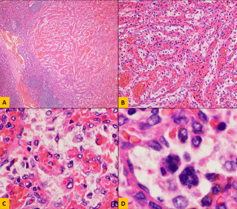
Answer: D. Littoral Cell Angioma
Discussion:
Littoral cell angioma is a benign vascular neoplasm of red pulp lining cells that is often multifocal. Lining cells have features of histiocytes (CD163+) and endothelial cells (CD31+, CD34+/-). Extramedullary hematopoiesis may be present (megakaryocytes are the most obvious evidence of this, seen in image D of case images). The top differential diagnoses are Hemangioma (endothelial lining cells only with no histiocytic features), Angiosarcoma (more atypia, interanastomosing vascular channels, necrosis, and mitoses), Littoral Cell Angiosarcoma (rare, lining cells are atypical), hamartoma (CD34, CD31, and factor VIII are negative except in incidental vessels), hemangioendothelioma (more atypia), and Kaposi Sarcoma (HHV8+).
Source: ExpertPath, Littoral Cell Angioma, Nadine Aguilera, MD.
Case contributed by: Diana Morlote, M.D., Assistant Professor, Genomic Diagnostics and Bioinformatics