Case History
28-year-old male, zygoma mass, diagnosis?
- LG fibromyxoid sarcoma
- Chondromyxoid fibroma
- Chondrosarcoma
- Cellular myxoma
Answer: B.) Chondromyxoid fibroma
Discussion:
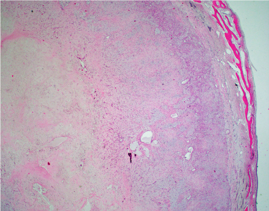
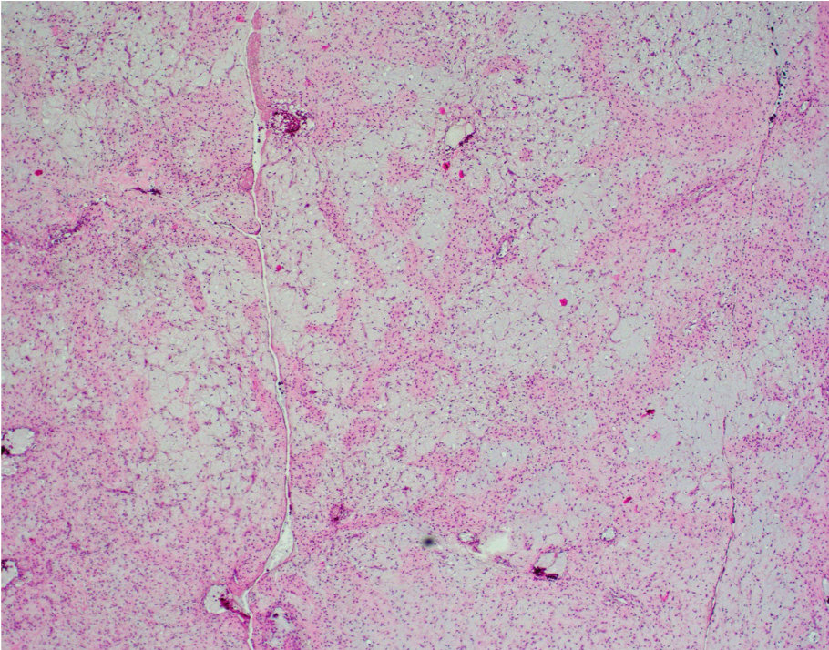
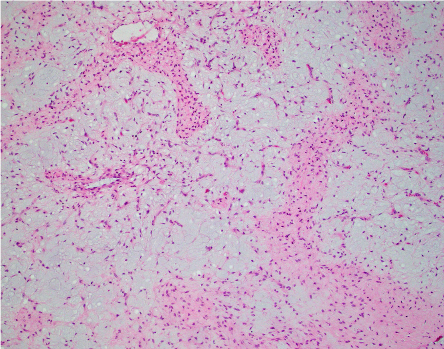
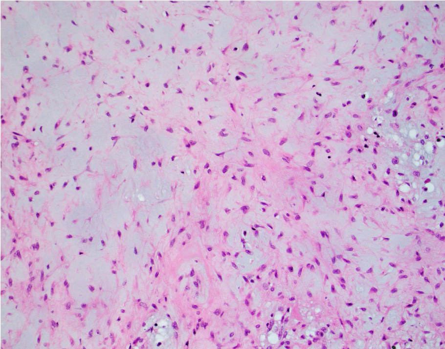
Chondromyxoid fibroma (CMF) is a benign neoplasm sharply demarcated from the surrounding bone exhibiting a zonal/lobular architecture. The peripheries of the lobule are more hypercellular and exhibit stellate and spindle-shaped cells and occasional osteoclast-like giant cells. The centers of the lobule are more hypocellular and myxoid with embedded bland stellate and spindle-shaped cells. While the centers of the lobule resemble hyaline cartilage, true hyaline cartilage is rare in CMF.
CMF may occur at almost any osseous site, most frequently in the long bones around the metaphyseal region.
WHO Classification of Soft Tissue and Bone Tumours. 5th ed.
Case contributed by: Todd Stevens, M.D., Associate Professor, Anatomic Pathology, Section Head - Head & Neck