Case History
A 22-year-old P0 woman presents in active labor at term, with subsequent uncomplicated vaginal delivery. Neonatal Apgar scores of 8 and 9. Mother had no prenatal care. Placenta sent to pathology to evaluate an attached mass.
What is the diagnosis?
- Accessory placental lobe
- Chorangioma
- Chronic hemorrhage
- Fetus papyraceus
Answer: D, Fetus papyraceus
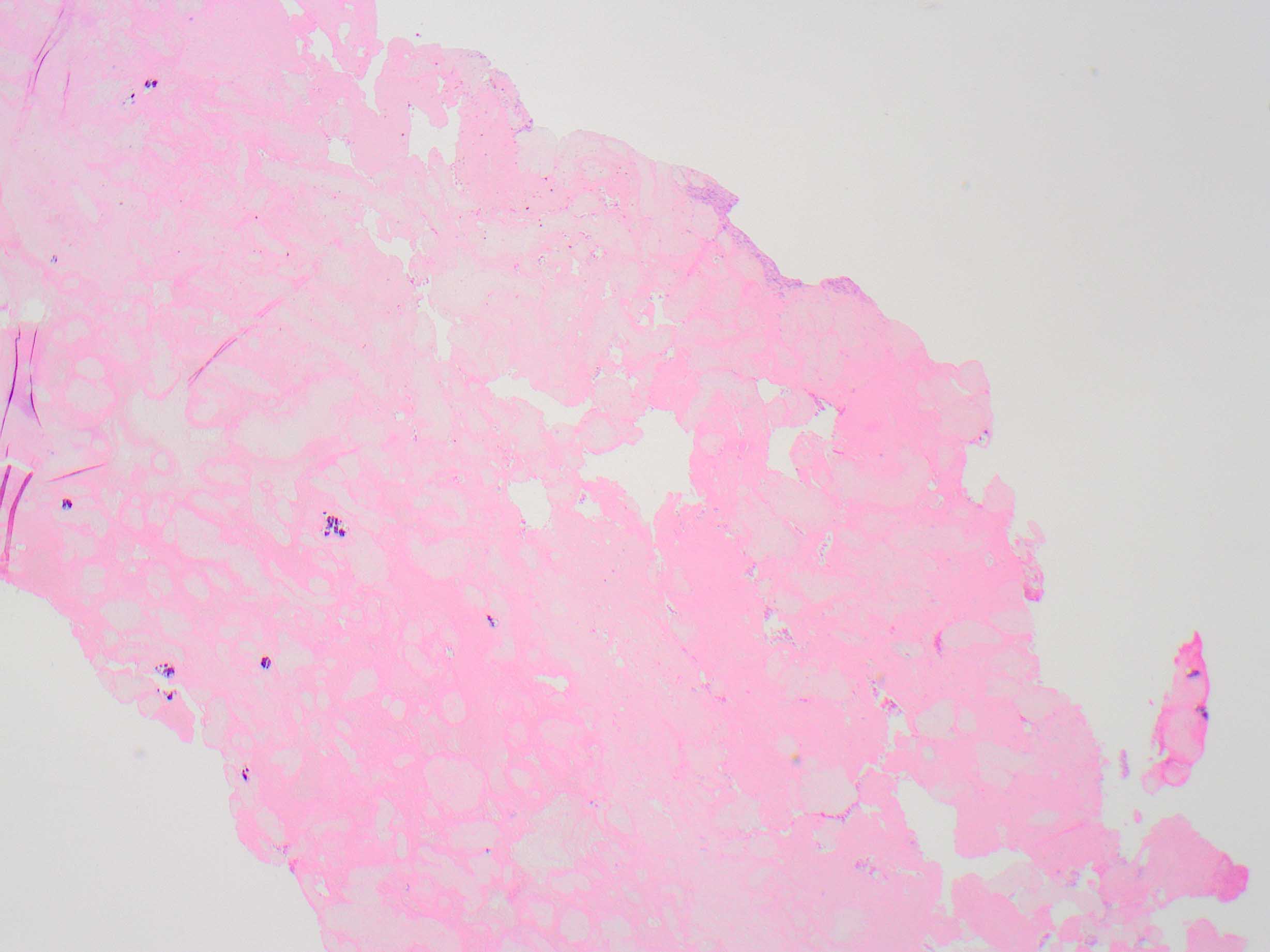
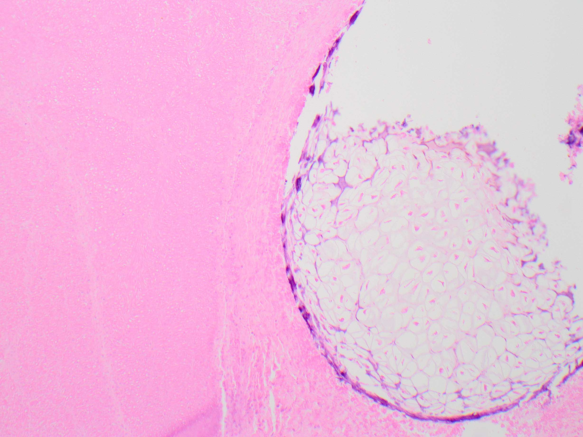
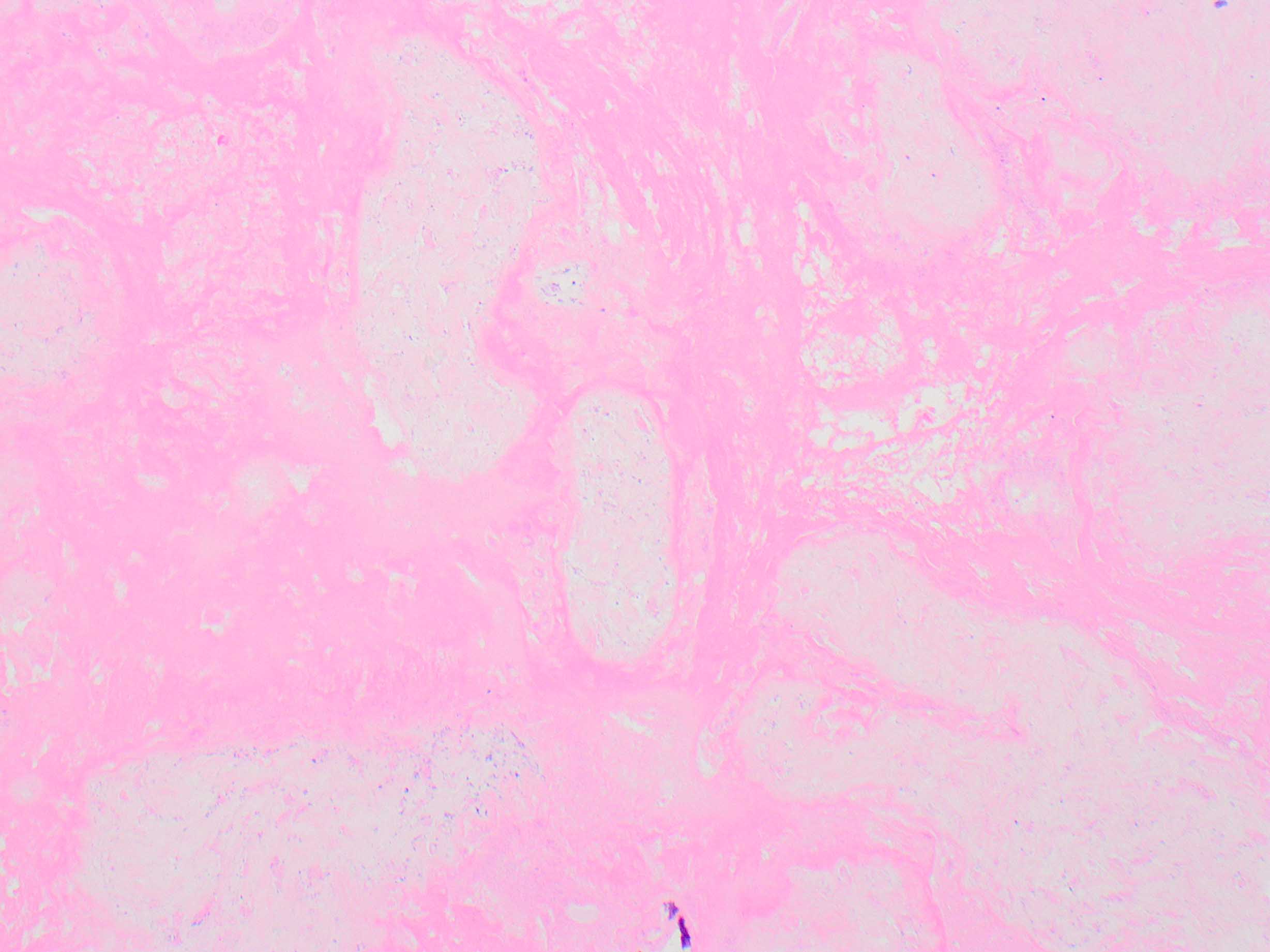
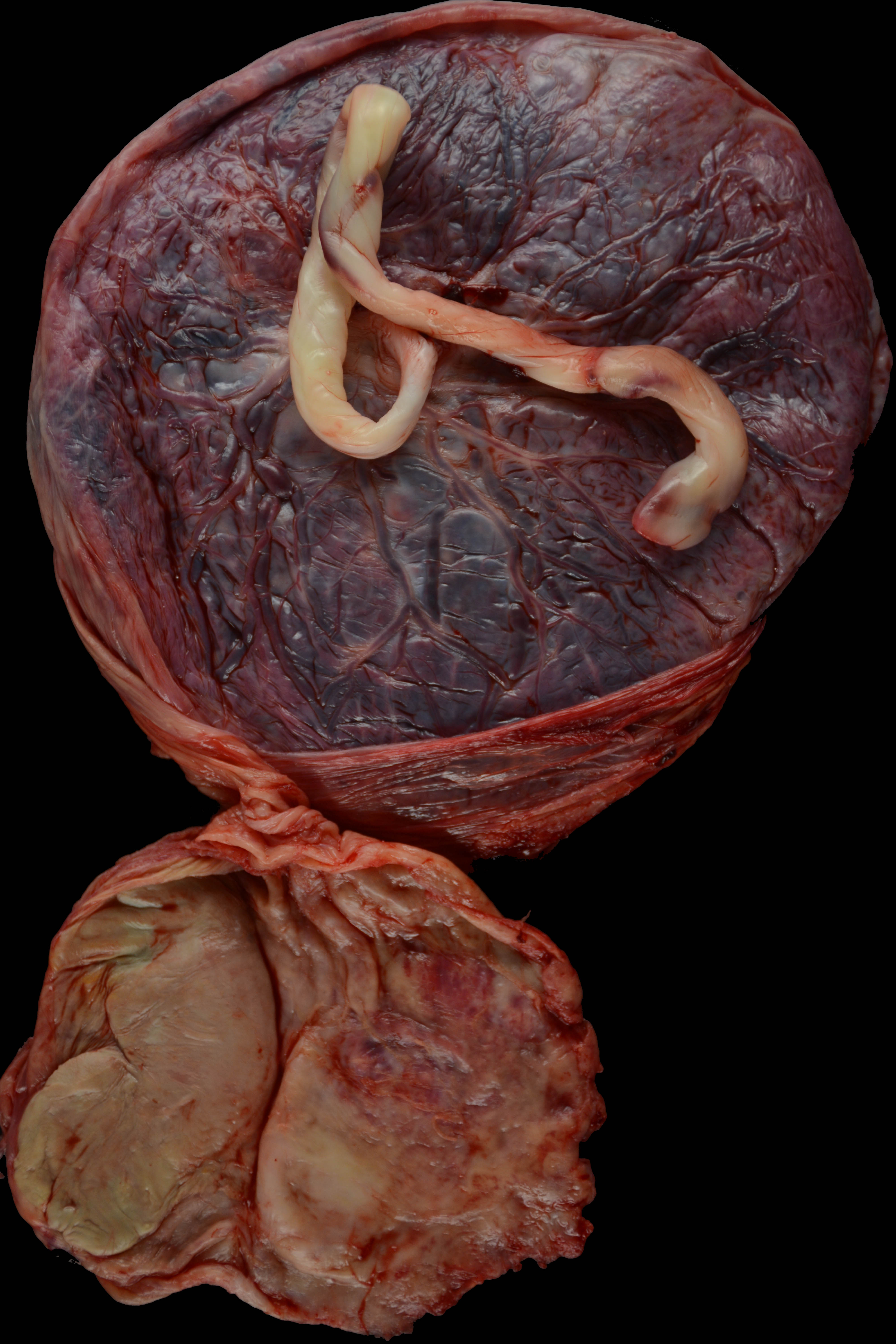
Gross examination of the placental specimen revealed a normal term placenta with a firm, ovoid mass immediately adjacent to the placental margin, as shown in the gross photograph of the fetal surface. The smaller mass was separated from the main placenta by an opaque membrane histologically confirmed to represent a diamniotic, dichorionic “dividing” intertwin membrane. Careful removal of the membranes overlying the mass showed a smooth ovoid form with vague suggestion of crown and rump regions, firmly affixed to a completely involuted apparent placental disc. Putative crown-rump measurement approximated 12-13 weeks of gestation at time of demise. Photomicrographs confirmed autolytic developing hyaline cartilage and adjacent muscle bundles, and completely involuted chorionic villi in the placental disc. The findings are diagnostic of fetus papyraceous, or “vanishing twin.”
Fetus papyraceus occurs when there is loss of one or more fetuses in a twin or higher multiple pregnancy, with gradual compression and involution of the residual placental and fetal tissue. Early conceptuses may be inconspicuous within the membranes, and may be missed without careful inspection. Fetuses at later ages of gestation are more easily detected and evaluated.
Early antenatal imaging can identify twin gestation, and subsequent imaging can identify fetal loss. However, in patients who have limited prenatal care, or in situations where imaging is limited, fetal loss of one co-twin can be an unexpected finding. Careful dissection and evaluation may identify anomalies in the demised twin, with potential implications for the surviving twin. Fetal demise of a co-twin has also been associated with increased death of the co-twin, increase in neurodevelopmental abnormalities in the surviving twin, and skin defects in the surviving co-twin. These complications are particularly important in the case of monozygotic twins, and highlight the importance of a careful pathologic characterization of the fetus and membranes in these cases.
The differential diagnosis of the margin-associated placental mass in this case also includes an infarcted accessory placental lobe, chronic hemorrhage, and chorangioma. Accessory (succenturiate) placental lobe is a variation in placental shape consisting of a smaller lobe of placental tissue connected to the main mass by a membrane through which fetal vessels course to supply the accessory lobe. These may infarct and could have a gross appearance similar to the mass in this case, but would not include fetal tissue. Chronic hemorrhage may accumulate within the retromembranous and/or retroplacental space and form a tan-gray shaggy fibrinous mass that could at first glance be grossly mistaken for a fetus papyraceous, but closer inspection would reveal soft laminated to homogeneous tissue without placental or fetal tissue. Chorangioma is the most common placental tumor, thought to be hamartomatous, and are usually single and small. Rarely they can be quite large and involve the margin of the placenta or an accessory lobe, and these may be associated with fetal complications. However, the gross and microscopic appearances would differ from the mass in this case, and would not include placental or fetal tissue.
References
Benirschke K, Burton GB, Baergen R: Pathology of the human placenta (2012), 6th ed., Springer Verlag.
Livnat EJ, Burd L, Cadkin A, Keh P and Ward AB: Fetus papyraceus in twin pregnancy. Obstet Gynecol 1978;51(1 Suppl):41s-45s
Pieretti ML, Alcalá R, Boggio P, Noguera-Morel L, Porriño ML, Luna PC, Hernández-Martín A, Schroh R, Larralde M, Torrelo A. Aplasia Cutis Congenita Associated with Fetus Papyraceus. Pediatr Dermatol. 2015 Nov-Dec;32(6):858-61.
Yoshida K, Soma H. Outcome of the surviving cotwin of a fetus papyraceus or of a dead fetus. Acta Genet Med Gemellol (Roma). 1986;35(1-2):91-8.