Case History
This is a 65-year-old man with a remote history of CVA, discovered to have a left atrial mass by echocardiogram following a syncopal episode.
What is the diagnosis?
- Organizing thrombus
- Papillary fibroelastoma
- Cardiac myxoma
- Hemangioma
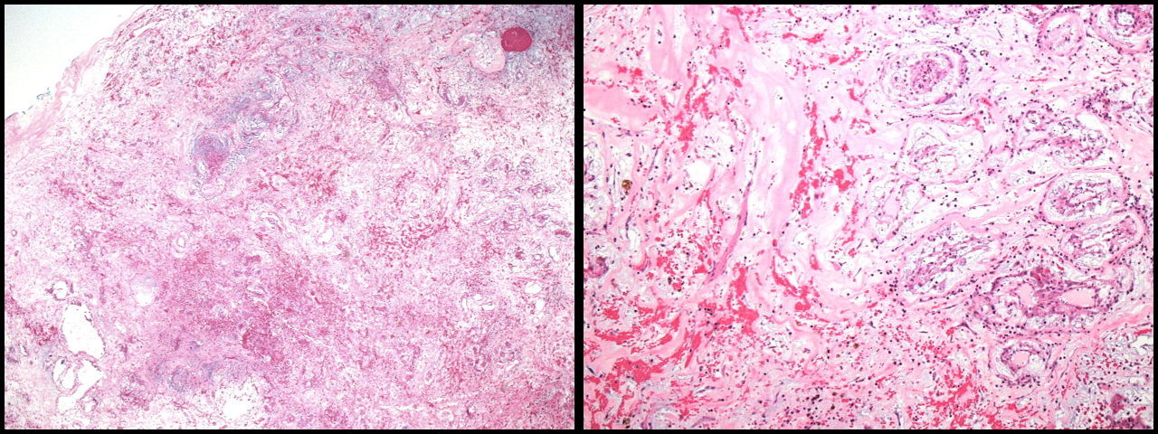
The answer is “C”, cardiac myxoma (Example “A”, Granular cell tumor)
Cardiac myxomas arise from the endocardium, most frequently in the atria and in the vicinity of the fossa ovalis however they can arise from other areas of the endocardium. They do not typically involve the valves. The histologic appearance of atrial myxoma is characteristic but not uniform. The tumor is composed of varying amounts of myxoid stroma admixed with edematous and fibrous areas. Hemorrhage both fresh and old are typically present. Calcification can be seen. A vascular proliferation is always present and the vessels are patent with an edematous ring surrounding the lumen. Perivascular hyalinization is frequently seen. The tumor cell is dendritic in architecture. The surface is frequently covered in thrombus and in the past some pathologists considered myxomas to be a manifestation of organizing thrombi. Papillary fibroelastoma occurs on the valve surface and has an arborizing architecture. It can be associated with thrombus that can be confused with myxoma. Cardiac hemangiomas are rare but can be confused with myxoma due to the vascular proliferation. Carney complex, consisting of skin lesions, myxomas of the heart and skin, schwannomas and endocrine disorders accounts for a small proportion of cardiac myxomas.
References
Jagdish Butany, and L Maximilian Buja. Cardiovascular Pathology. London, Elsevier Academic Press, 2016. 748-753
Vindhyal MR, Elshimy G, Elhomsy G. Carney Complex. 2020 Jul 17. In: StatPearls [Internet]. Treasure Island (FL): StatPearls Publishing; 2020 Jan