Case History
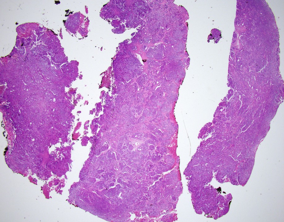
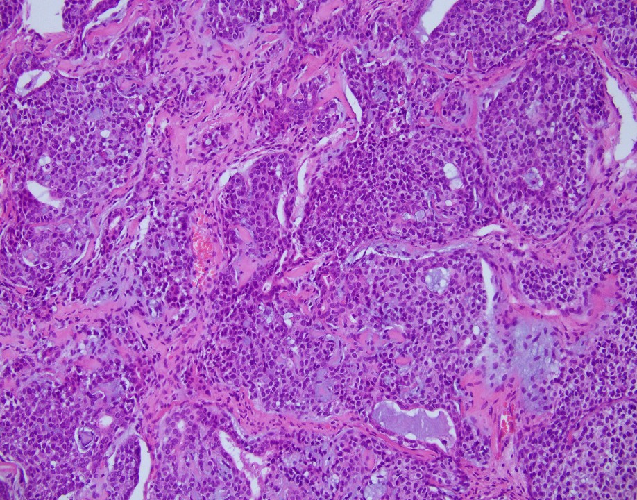
75 years old female. MR image shows a 5.2 x 4.4 x 4.0 cm perianal mass. This is left perirectal mass biopsy:
What is the diagnosis?
- Adenoid cystic carcinoma
- Benign cellular mixed tumor
- Malignant mixed tumor
Answer: A. Adenoid cystic carcinoma
The neoplasm reveals mixed epithelial and myoepithelial components, without significant cytologic atypia, embedded in a chondromyxoid stroma. The epithelial component shows strong cytoplasmic positivity with cytokeratin (AE1/AE3), while the myoepithelial component is positive for S100 protein and p63 (with all appropriate controls). Salivary Duct Tumor Gene Fusion Analysis detected MYB-NFIB gene fusion, which supports this is an adenoid cystic carcinoma.
Case contributed by: Shoujun Chen, M.D., Ph.D., Breast/GYN Fellow