Case History
A 52-year-old female presented with a breast mass.
A lumpectomy specimen shows the lesion illustrated in the images (A-D).
By IHC, what immunophenotypic profile this lesion is most likely to express?
-
A. ER+/PR+/HER2-
B. ER+/PR+/HER2+
C. ER-/PR-/HER2+
D. ER-/PR-/HER2-
Answer D. ER-/PR-/HER2-
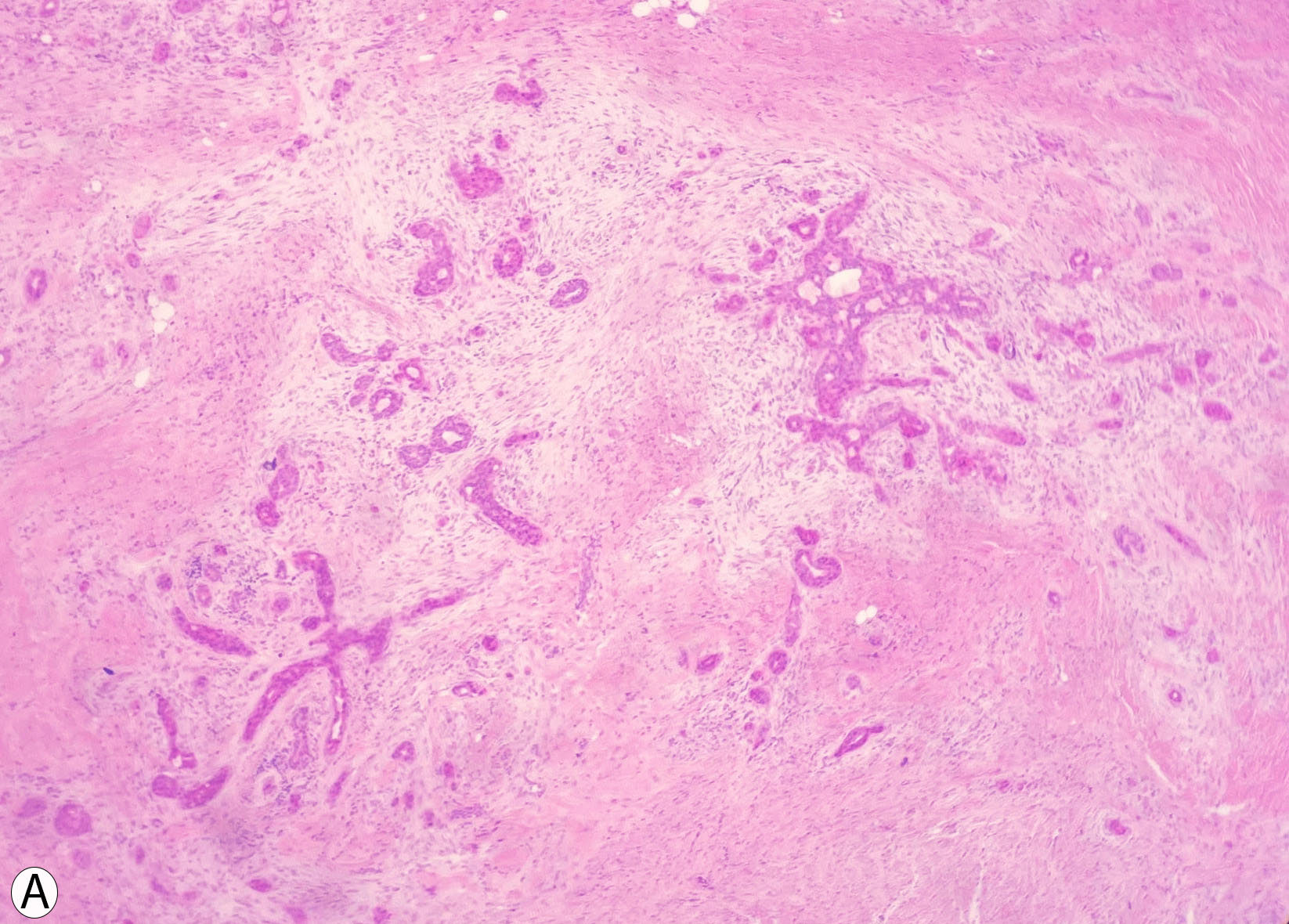
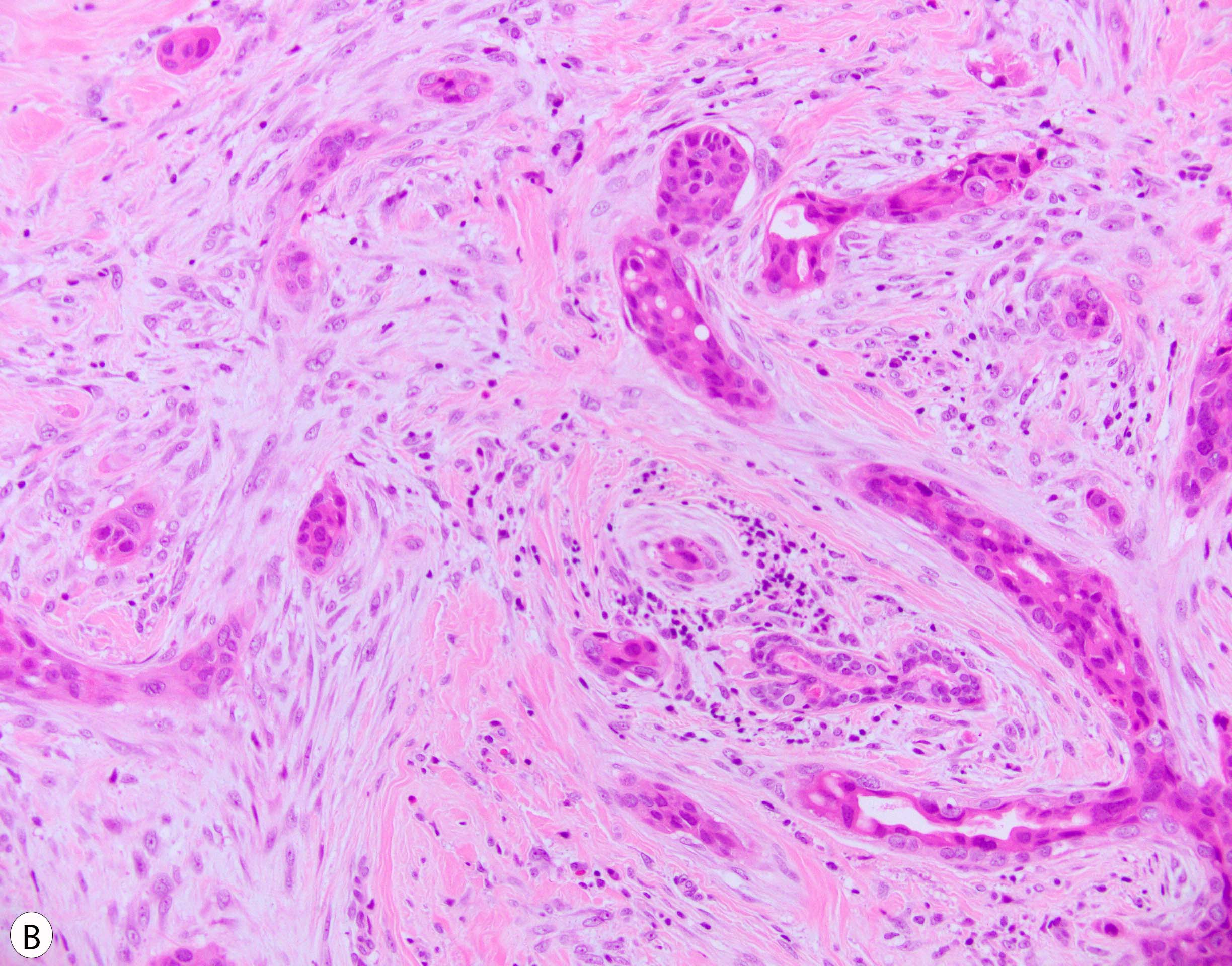
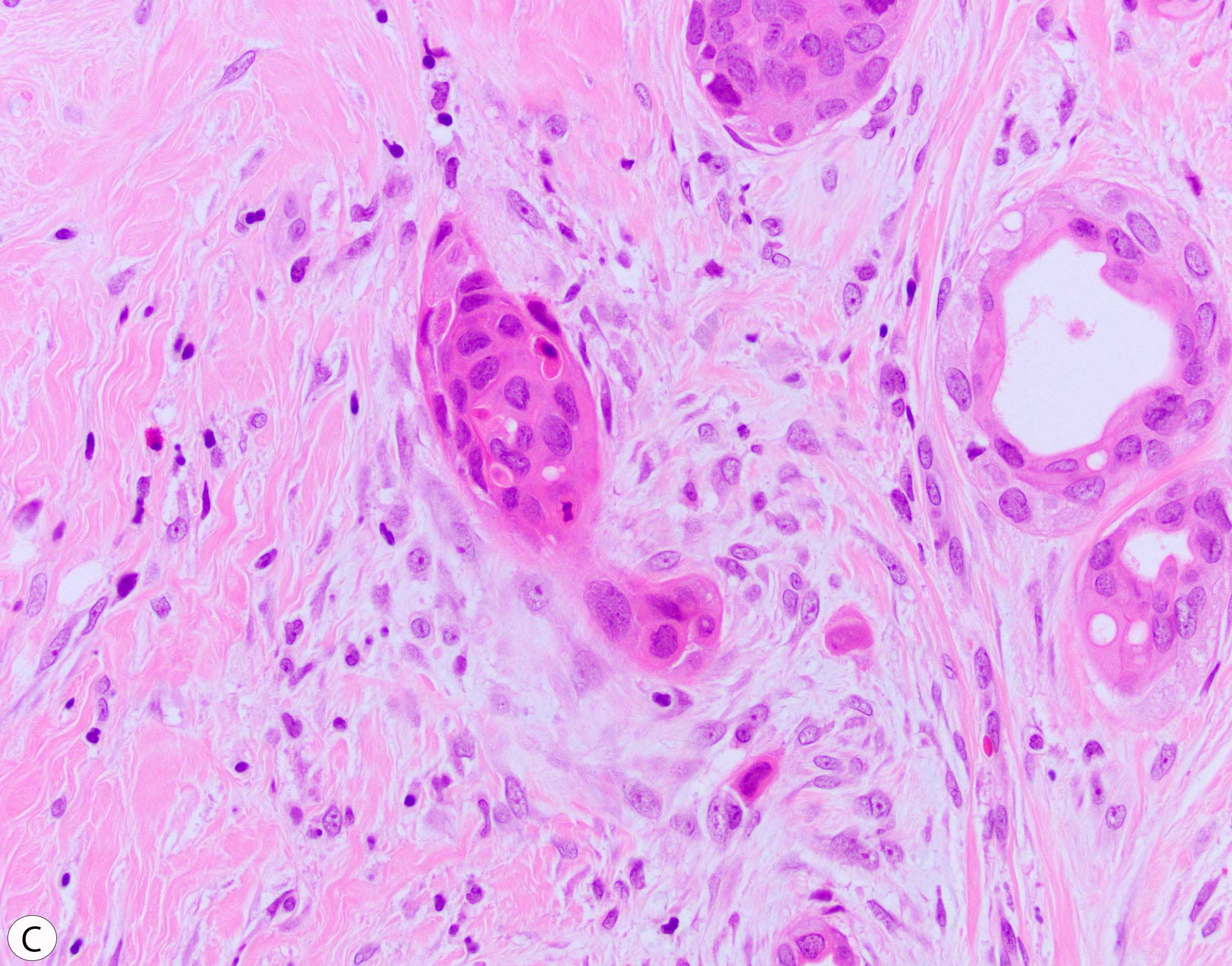
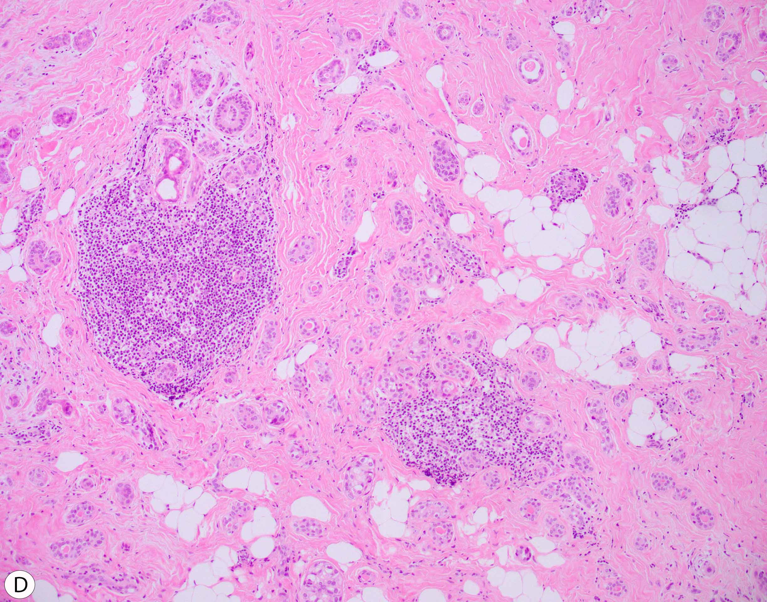
Low-grade adenosquamous carcinoma (LGASC) is a rare form of metaplastic breast carcinoma (MBC) characterized as a tumor with both glandular and squamous differentiation in a background of spindle cell proliferation (figure A). The glandular component is composed of multiple layers with low nuclear grade (figure B) that may resemble radial scar, tubular carcinoma, and syringomatous tumor of the nipple. The squamous component typically shows foci or nests of squamous cells, sometimes in “comma shape”, with keratin pearls (figure C). The surrounding spindle cells represent neoplastic proliferation with bland cytomorphology that usually merge with the adjacent epithelial components. Peripheral clusters of lymphocytes in a “cannonball pattern” may also be seen (figure D).
Immunohistochemical staining is not required for the diagnosis. However, if used, the glandular component shows a variable degree of positive expression for luminal and basal cytokeratins, with p63 expressed in the outer cell layer (but other myoepithelial markers are negative), as well as in squamous cells. Similarly, the spindle cells may show variable positive expression to cytokeratins and p63.
More than 90% of MBCs including LGASC are triple-negative by immunohistochemical study, whereas protein expression profiling shows that MBC is a heterogeneous group of tumors that can be classified as basal-like, claudin-low, and/or mesenchymal-like cancers.
In contrast to other forms of MBC, LGASC has a favorable prognosis. The latter might be attributed to high rates of PIK3CA mutation detected in LGASC and lack of TP53 mutation that is frequently seen in other forms of MBC.
Selected references:
WHO Classification of Tumours: Breast Tumours. 5th ed. International Agency for Research on Cancer, 2019
Geyer FC, Pareja F, Weigelt B, et al. The Spectrum of Triple-Negative Breast Disease: High- and Low-Grade Lesions. Am J Pathol. 2017;187(10):2139-2151. doi:10.1016/j.ajpath.2017.03.016
Bataillon G, Fuhrmann L, Girard E, et al. High rate of PIK3CA mutations but no TP53 mutations in low-grade adenosquamous carcinoma of the breast. Histopathology. 2018;73(2):273-283. doi:10.1111/his.13514