Case History
72 years female with a history of acral melanoma presented with loss of appetite, a mass in pancreatic head and multiple small nodules in peripancreatic fat. Endoscopic ultrasound fine needle aspiration was performed.
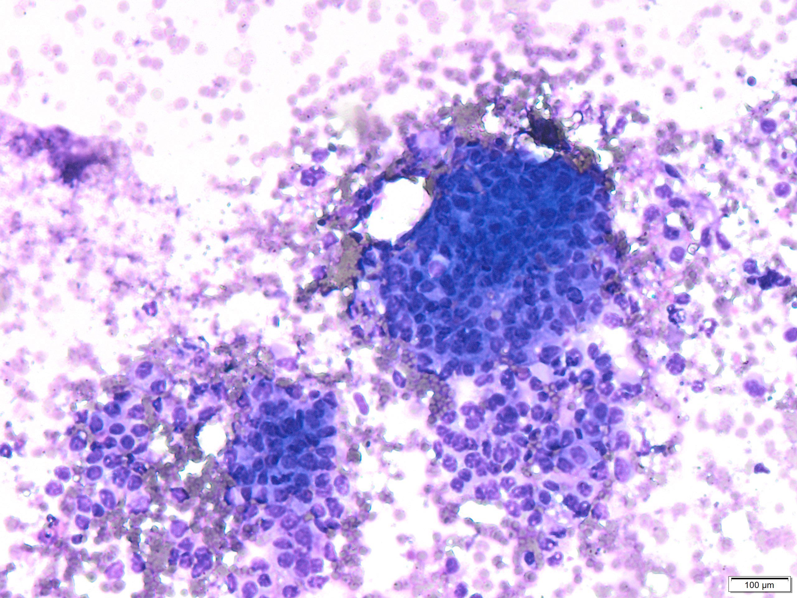
- Based on Diff-Quick stain smears rank the following diagnosis from most likely to unlikely:
- -Solid pseudopapillary carcinoma
- -Carcinoid tumor
- -Islet cell tumor
- -Acinar carcinoma
- -Poorly differentiated ductal carcinoma
- Positive staining for neuroendocrine markers likely exclude (mark true or false):
- -Solid pseudopapillary carcinoma
- -Carcinoid tumor
- -Islet cell tumor
- -Acinar carcinoma
- -Poorly differentiated ductal carcinoma
- Although not frequently used, electron microscopy can differentiate between (mark true or false):
- -Insulinoma and carcinoid
- -Acinar carcinoma and ductal carcinoma
- -Solid pseudopapillary carcinoma and Islet cell tumor
- -Acinar carcinoma and carcinoid
- -Metastatic melanoma and poorly differentiated carcinoma
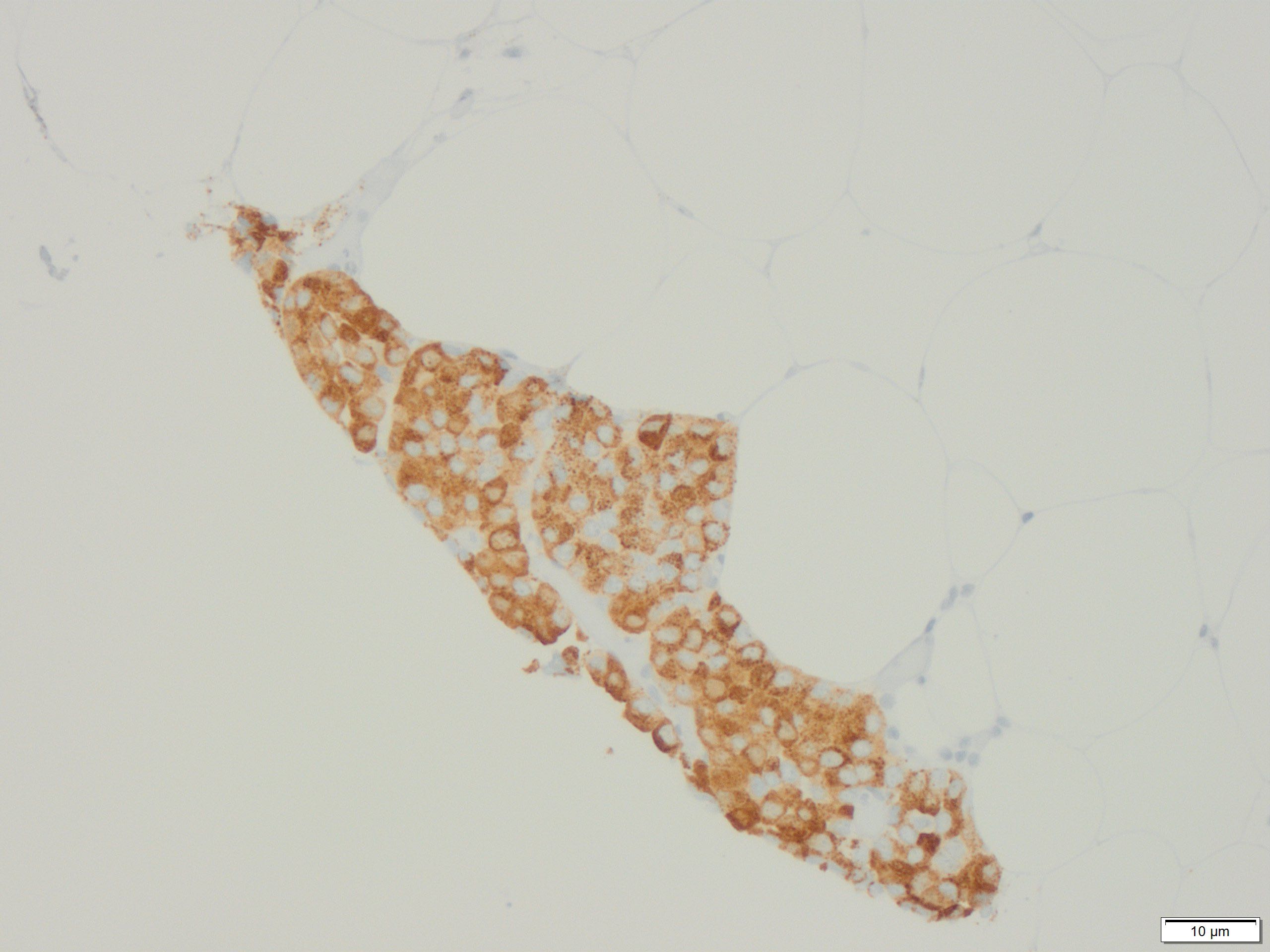 IHC positive staining for trypsin
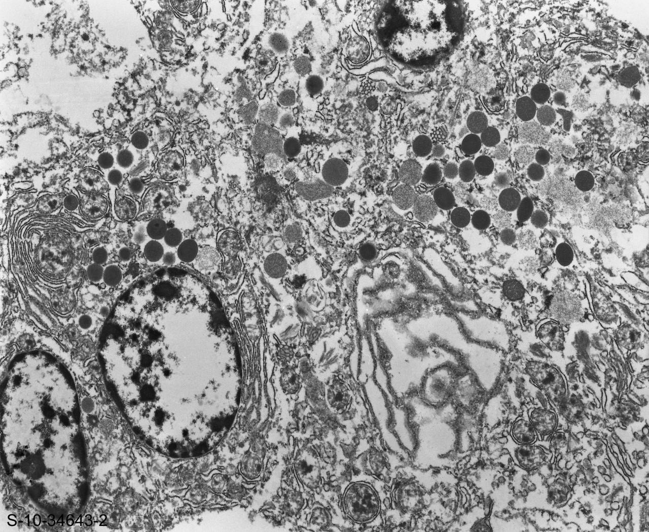
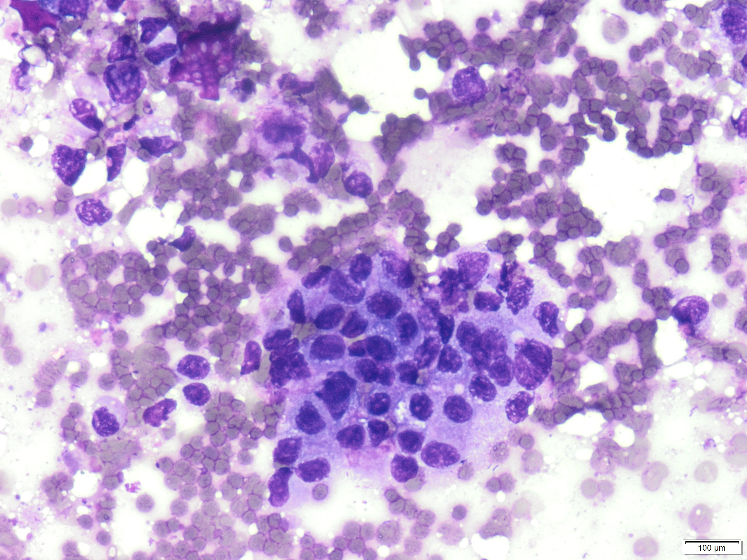
Electron microscopy image showing zymogen granules 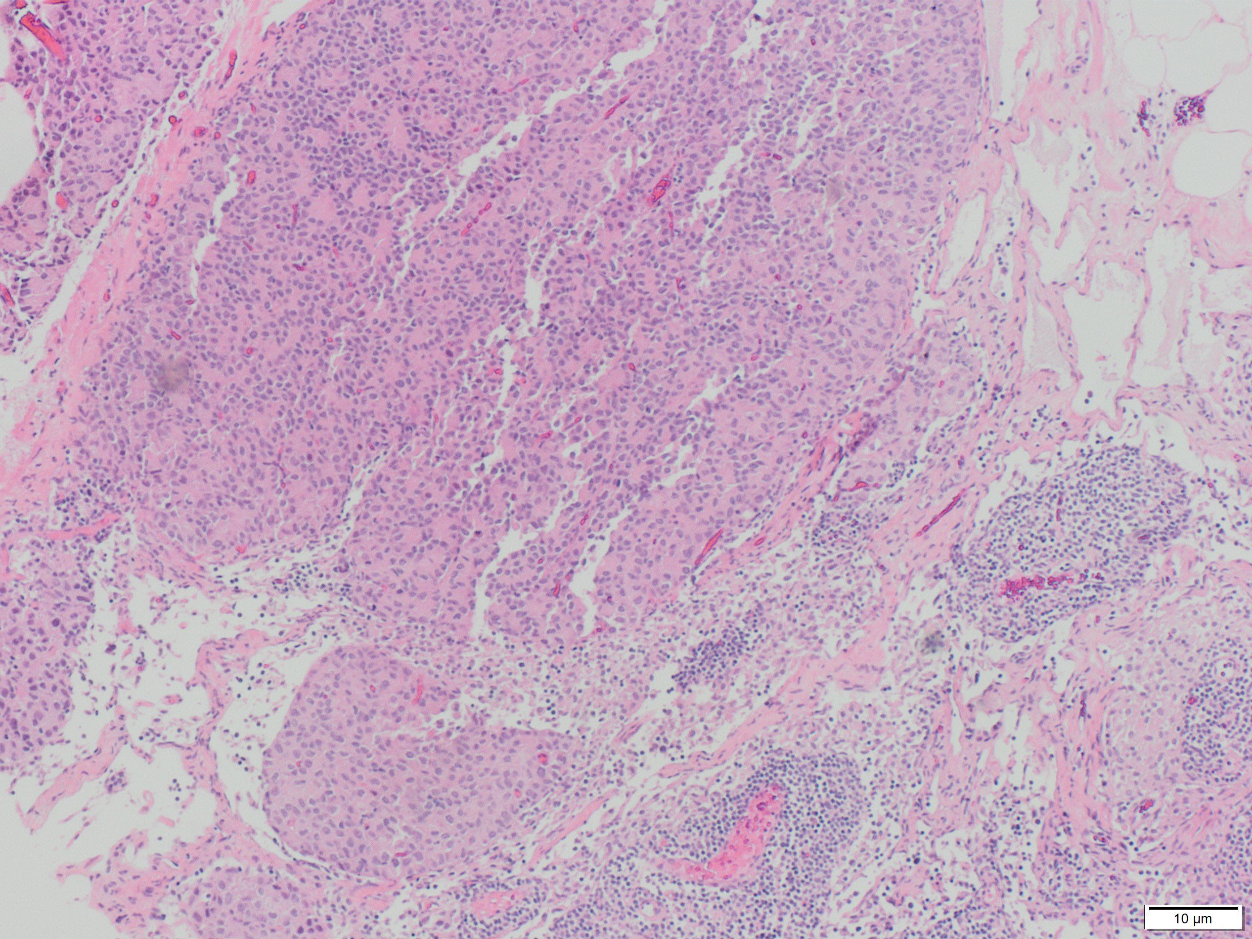 H&E-stained section showing metastatic acinar carcinoma in lymph node
-
Answers:
1:
1- Acinic carcinoma
2- Carcinoid tumor
3- Islet cell tumor
4- SPN
5- Poorly differentiated carcinoma -
2:
1- False
2- False
3- False
4- False
5- True -
3:
1- True
2- True
3-True
4-True
5-True -
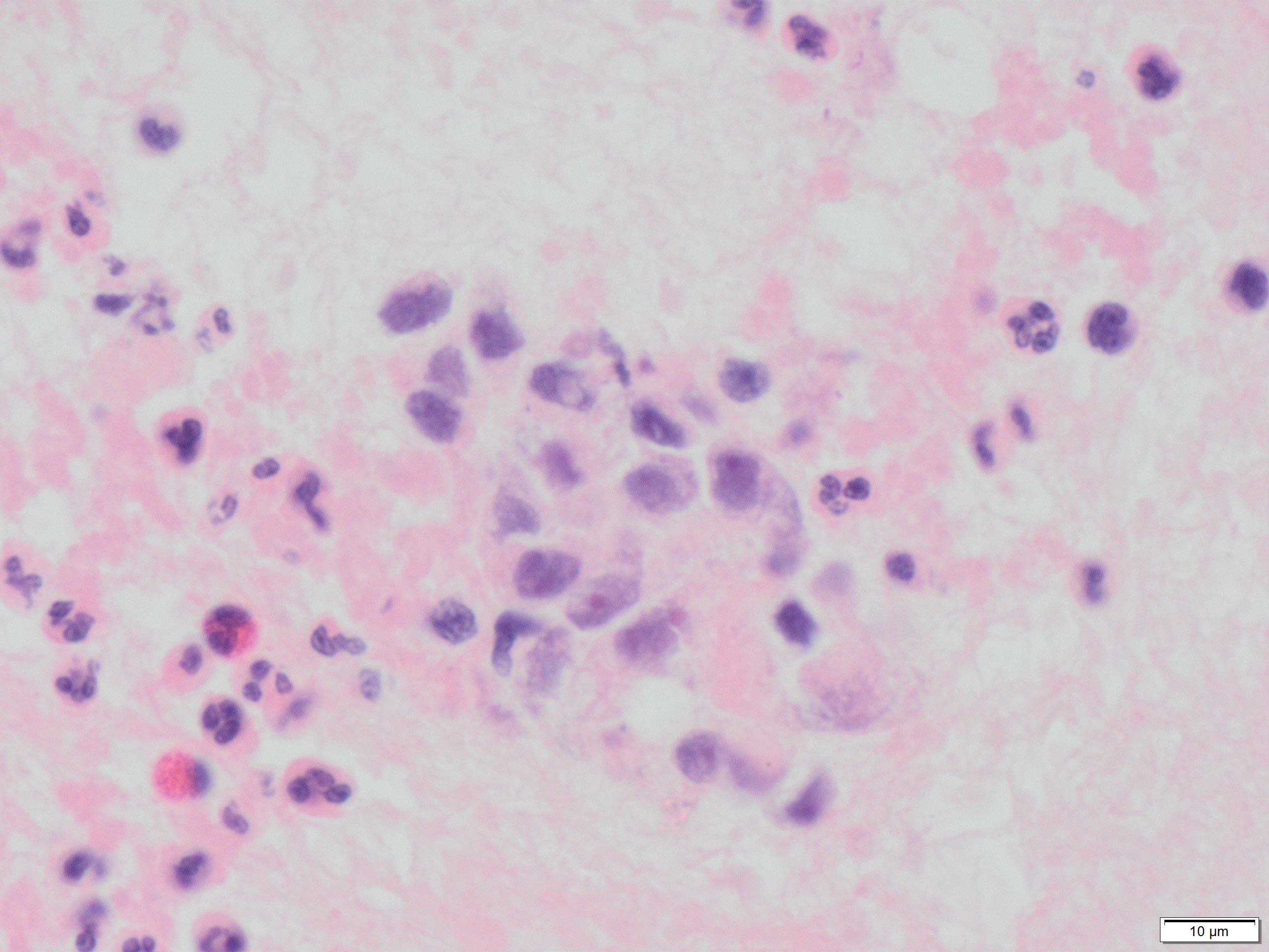
Stains for endocrine markers were negative. Stain for trypsin was positive. Electron microscopic examination shows classical zymogen granules. Final diagnosis acinic carcinoma with metastasis to lymph nodes.