Case History
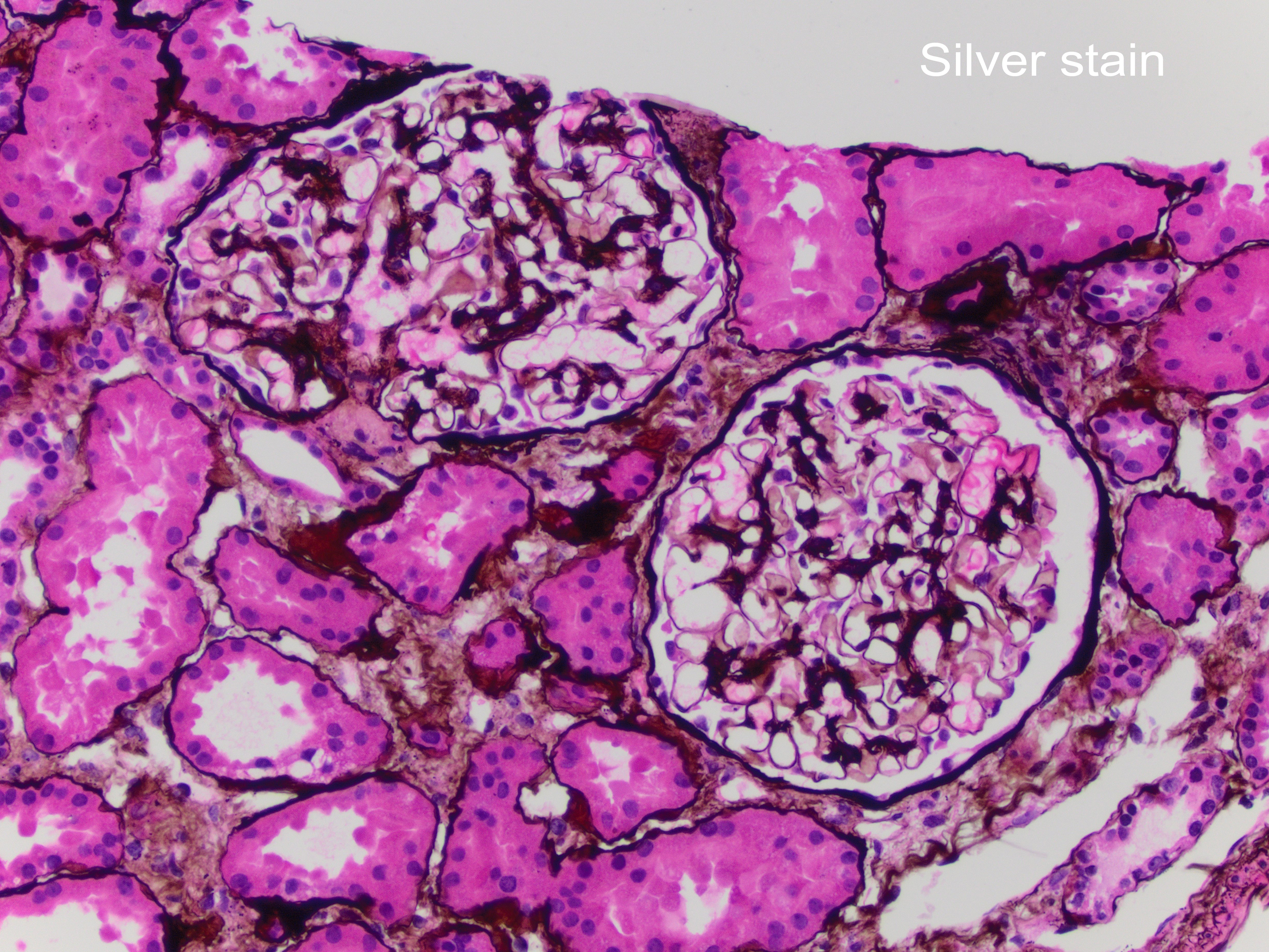
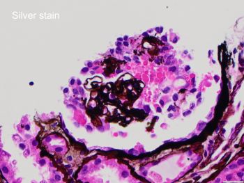
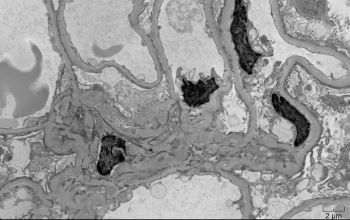
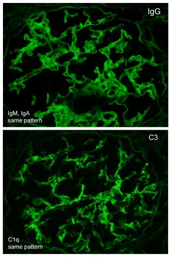
A 42-year-old African-American female with a history of systemic lupus erythematosus with acute kidney injury (creatinine 2.7 mg/dL), proteinuria (1064 mg/24 hr), and no hematuria, was admitted for lupus encephalitis. Renal biopsy by light microscopy showed that most glomeruli had no lesions (Figure 1) with one glomerulus with glomerular tuft collapse, and podocyte hypertrophy/hyperplasia (Figure 2). There was positive mesangial staining by immunofluorescence with IgG, IgM, IgA, C3, C1q, and light chains (Figure 3). Electron microscopy findings included diffuse foot process effacement, and mesangial electron-dense deposits (Figure 4).
What gene variants are strongly associated with these findings?
A. WDR73
B. PDSS2
C. APOL1
D. SCARB2
Case contributed by: Andrea Kahn, M.D., Professor, Anatomic Pathology, GYN Section Head