Case History
A 7 y/o M with pancytopenia and splenomegaly. Which one of the following is NOT included in DDx:
- Gaucher disease.
- MAC infection.
- Mast cell disorder
- Whipple disease
Answer: C. Mast cell disorder
Discussion
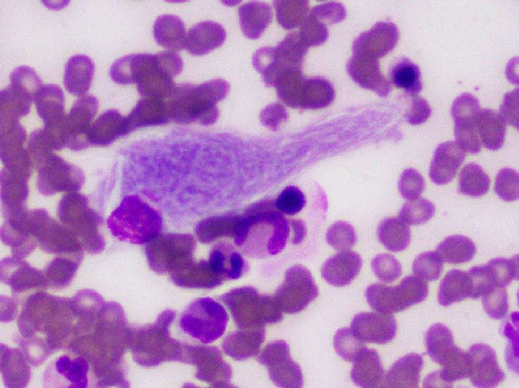
This is a case of Gaucher disease (A). Mycobacterium avium complex (MAC) infection (B) and Whipple disease (D) are included in the differential diagnosis due to similar morphologic picture of clustering histiocytes with so-called “wrinkled paper-like” cytoplasm. Atypical mast cells are larger than basophils with irregular, elongated spindle shapes or ovoid shapes. Their cytoplasm is packed with basophilic granules that may obscure nuclear margin and the nucleus is round and single.
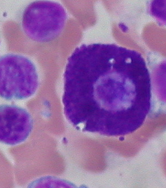 Mast cell
Autosomal recessive disease due to accumulation of glucocerebroside / glucosylceramine (a sphingolipid) in reticuloendothelial cells in liver, spleen and bone marrow, due to a defect in lysosomal beta glucocerebrosidase.
Increased risk of 14x for hematologic malignancies and 4x for other malignancies.
Diagnosis
Absence of glucocerebrosidase in peripheral blood monocytes.
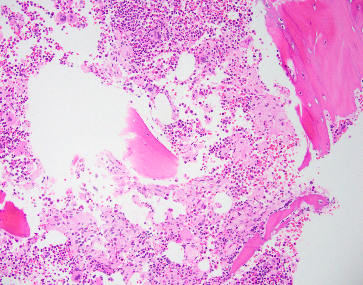
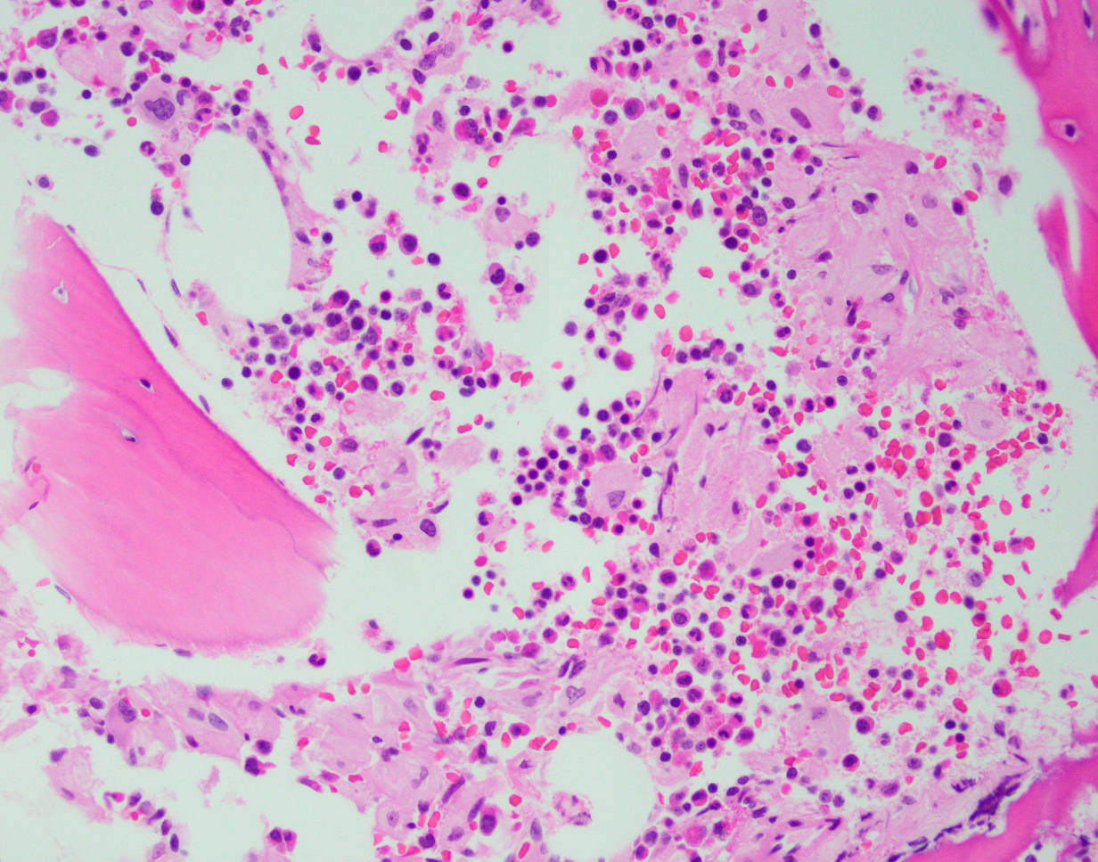
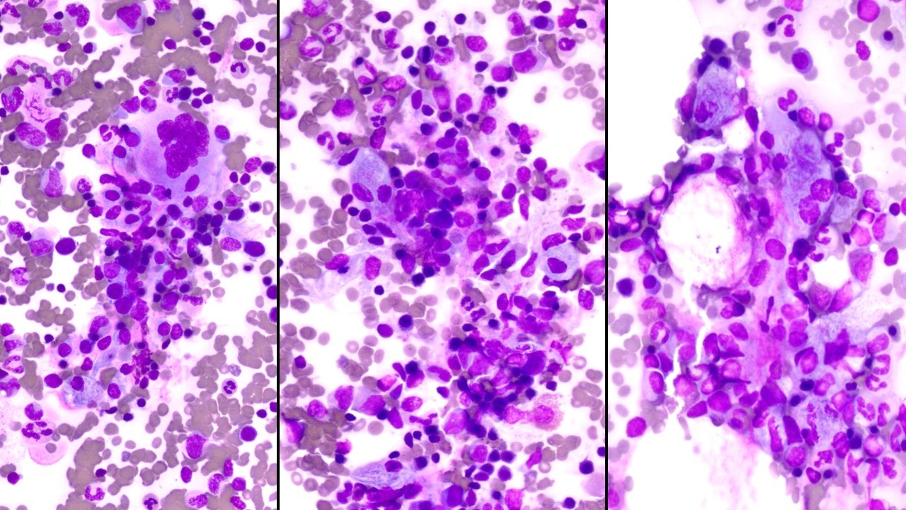
Microscopic description
Small focal accumulations or diffuse replacement by ovoid histiocytes 20 - 90 microns, with abundant, finely fibrillar, pale blue gray cytoplasm that is crinkled or wrinkled paper-like
Small nucleus with coarse chromatin and indistinct nucleolus
Increase in reticulin fibers
Stains
Positive: Iron, CD68, PAS, TRAP, NSE
Negative: Sudan Black B, AFB
Differential diagnosis
Chronic myeloid leukemia
Histiocytic disorders
Lipid storage diseases (e.g. Niemann Pick disease) (PAS-, Sudan Black B+)
Mycobacterium avium complex (MAC) infection (PAS+, AFB+)
Whipple disease (PAS+, AFB-)