Case History
A 75-year-old female with limited past medical history presented from an outside hospital for endoscopic resection of a gastric mass.
What stain would be the most helpful in determining the diagnosis?
A. Pancytokeratin
B. GATA3
C. ERG
D. CK7
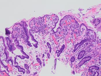
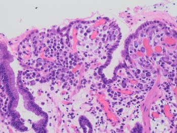
As shown on the H&E, the gastric mucosa shows atypical cells located predominantly in the superficial lamina propria. The atypical cells are small, discohesive cells with targetoid mucin, minimal nuclear atypia without stromal desmoplasia or necrosis. The background mucosa shows mild reactive changes but no intestinal metaplasia.
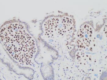
Given the lack of clinical history and the tumor cell location in the superficial gastric pits, the main differential diagnosis’ include diffuse-type gastric carcinoma and metastatic lobular carcinoma. As noted in the question stem, limited clinical history is provided, therefore a prior history of breast cancer is unknown. Therefore, GATA3 stain (SHOWN HERE) shows strong and diffuse staining in the tumor cells, thus supporting a tumor of breast origin (i.e. lobular carcinoma). In addition, The tumor was positive for ER, further supporting a metastatic lobular carcinoma.
Pancytokeratin would stain these tumor cells and establish epithelial origin, but would not further differentiate the tumor. ERG would stain vascular cells, which would not be positive in these cells. CK7 would stain in both gastric and breast carcinomas, so it would not further differentiate the tumor. Therefore, GATA3 is the best answer choice.
Contributed by Chirag Patel, M.D., Assistant Professor, Anatomic Pathology