Case History
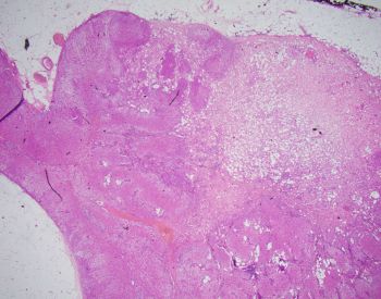
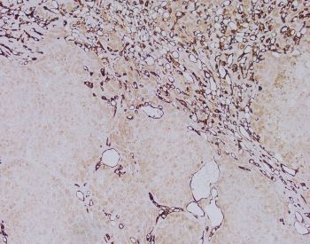
A 48-year-old man presents with an incidental 3 cm right adrenal mass, undergoes right adrenalectomy. The tubular and canalicular structures are positive for calretinin.
- Adrenal cortical adenoma
- Metastatic signet ring cell carcinoma
- Adenomatoid tumor
- Composite adrenal cortical adenoma and adenomatoid tumor
Discussion:
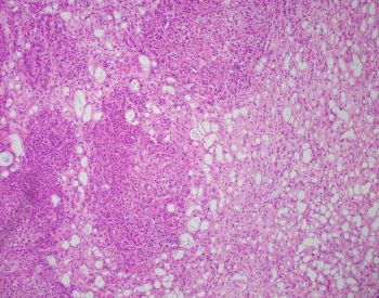
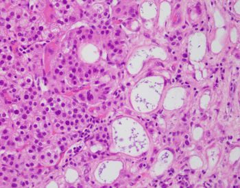
On histologic examination, this nodule consisted of adrenal cortical adenoma admixed with canalicular and tubular (gland like) structures. Tubular structures were typically characterized by flat cells. Some of the cells exhibited vacuolated cytoplasm resembling signet ring cells.
The tubular and canalicular structures were positive for calretinin, and adrenal cortical adenoma was negative for calretinin.
Adenomatoid tumor is a benign neoplasm of mesothelial origin that is generally seen in paratesticular adnexa, uterus and fallopian tubes. This entity has also been described in other organs including the adrenal gland.
Adenomatoid tumors are positive for mesothelial markers (calretinin, HBME-1), cytokeratins and thrombomodulin.
Complex tubules with gland like appearance are cell groups with round intracytoplasmic vacuoles, vesicular nucleus and small nucleolus may simulate metastatic adenocarcinoma or signet ring cell carcinoma.
In the present case, the lack of pleomorphism, necrosis and cellular atypia argue against malignancy.
In light of the morphologic and immunohistochemical findings, the diagnosis of composite adrenal cortical adenoma and adenomatoid tumor was rendered.