Case History
A 58-year-old woman presents with a new left upper quadrant breast mass. The tumor cells show SOX10 positivity.
What additional stain would help reach the diagnosis?
- Pancytokeratin; IDC grade 3
- P63; Metaplastic Carcinoma
- Melan-A; Metastatic Melanoma
- CK7; Adenoid Cystic Carcinoma
Correct Answer: C. Melan-A; Metastatic Melanoma
Discussion:
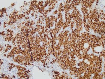
Sections show tumor cells exhibiting plasmacytoid morphology. The nuclei are pleomorphic and exhibit prominent nucleoli and fine granular pigment deposition. The histomorphology, in conjunction with the strong and diffuse SOX10 positivity, should raise suspicion for metastatic melanoma within the differential diagnosis. With the Melan-A stain also positive in the tumor cells, this lesion represents metastatic melanoma to the breast. Prior medical history of melanoma was confirmed on subsequent patient follow-up.
In addition to melanocytic cells, SOX10 stain can be used as a marker for breast myoepithelial cells and can stain positive in some triple-negative and metaplastic carcinomas. A tumor with pancytokeratin positivity would suggest a carcinoma, while p63 positivity would suggest a metaplastic carcinoma. If the tumor was bi-phasic (i.e. adenoid cystic), then the CK7 stain would highlight luminal cells while the SOX10 would highlight the basaloid cells. Given the Melan-A positivity, this lesion is consistent with metastatic melanoma.