Case History
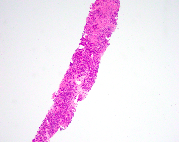
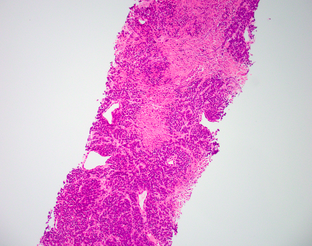
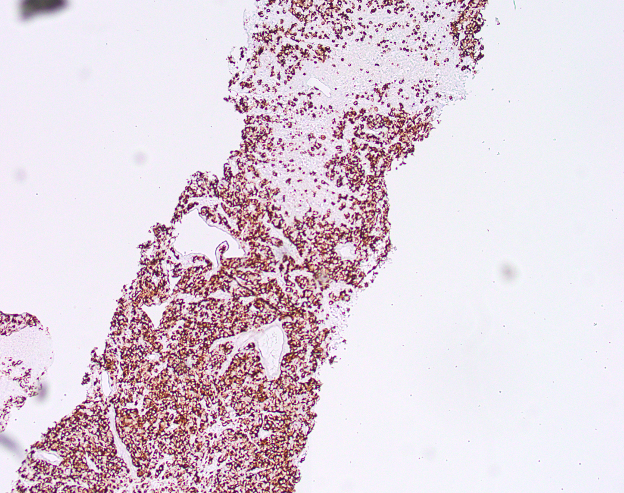
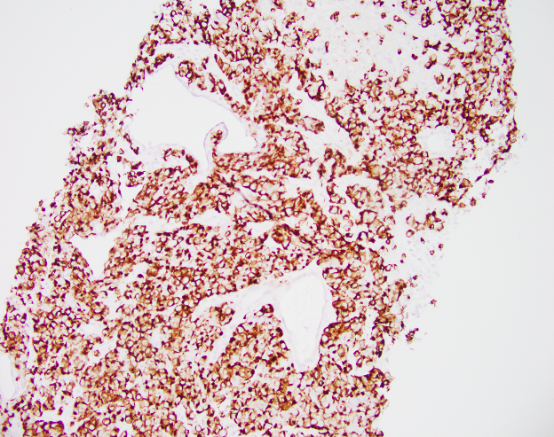
A 65-year-old male presents with left axillary lymphadenopathy. Sections show a small round blue cell tumor with high N:C ratio, finely dispersed chromatin, and indistinct nucleoli with apoptotic bodies and necrotic debris. Synaptophysin and chromogranin were both diffusely positive and CD 45 negative. Provided are the H&E and CK20 IHC images.
What is the best diagnosis?
- Ewing Sarcoma
- Lymphoma
- Merkel cell carcinoma
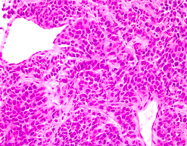
Correct Answer C. This is a case of a high grade neuroendocrine tumor, consistent with Merkel cell carcinoma.
Discussion
Merkel cell carcinoma is a highly aggressive primary cutaneous neuroendocrine carcinoma. There is emerging evidence of distinct Merkel cell polyomavirus association and UV mediated ongogenetic pathways. The classic dot-like paranuclear pattern is a distinctive feature of this entity as shown above.
References
1. Hodgson NC. Merkel cell carcinoma: changing incidence trends. Journal of surgical oncology. 2005; 89:1–4.
2. Feng H, Shuda M, Chang Y and Moore PS. Clonal integration of a polyomavirus in human Merkel cell carcinoma. Science. 2008; 319:1096–1100.
3. Harms PW, Vats P, Verhaegen ME, Robinson DR, Wu YM, Dhanasekaran SM, Palanisamy N, Siddiqui J, Cao X, Su F, Wang R, Xiao H, Kunju LP, et al. The Distinctive Mutational Spectra of Polyomavirus-Negative Merkel Cell Carcinoma. Cancer research. 2015.
Case contributed by: Raheel Ahmed, M.D. Surgical Pathology Fellow, UAB Pathology