Case History
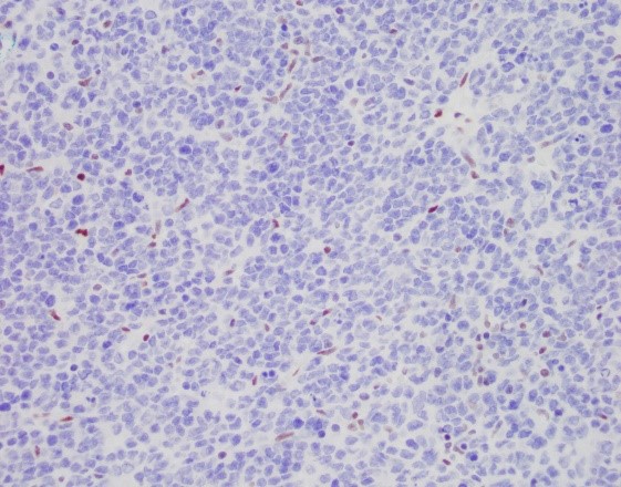
A 23-year-old female presents with CT identified 12 cm pelvic mass appearing to arise from right adnexa, suspicious for ovarian neoplasm. The ovary and fallopian tube were sent for pathology. Immunostaining result of BRG1 is provided.
What is the most likely diagnosis?
A. HGSC
B. SCCOHT
C. GCT
D. DLBCL
Discussion:
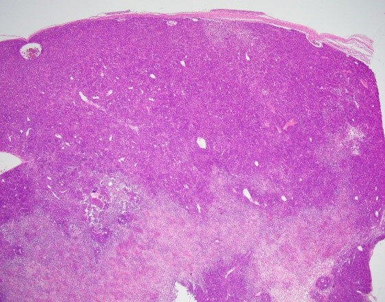
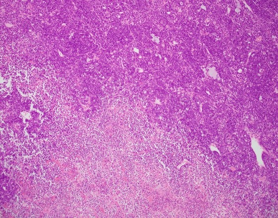
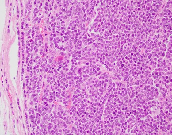
The sections show sheets of tumor cells in a background of marked necrosis. The tumor cells are small to intermediate in size, with scant cytoplasm, clump chromatin and small nucleoli. The tumor is positive for WT1, focally positive for CAM5.2, CD56, synaptophysin, negative for pan-cytokeratin, HMWCK, EMA, inhibin, OCT3/4, SALL4, CK7, CD10, chromogranin, SF1, and PAX8. P53 is wild type. CD45 highlights background lymphocytes. CD30 highlights scattered cells. P16 is non-contributory. SMARCA4/BRG1 staining shows absence of nuclear staining in the tumor cells (the nuclear stained stromal cells are used as internal control), supporting a diagnosis of small cell carcinoma of the ovary, hypercalcemic type.
Contributed by Dr. Junlin Zhang and Dr. Xiao Huang, UAB pathology