Case History
A 60 y/o female, prior smoker, with a 2x1 cm left parotid nodule noted 3 months prior.
What is the best diagnosis?
A. Tattoo
B. Pigmented schwannoma
C. Metastatic melanoma
D. Metastatic pigmented mammary carcinoma
The answer is C. metastatic melanoma.
Discussion:
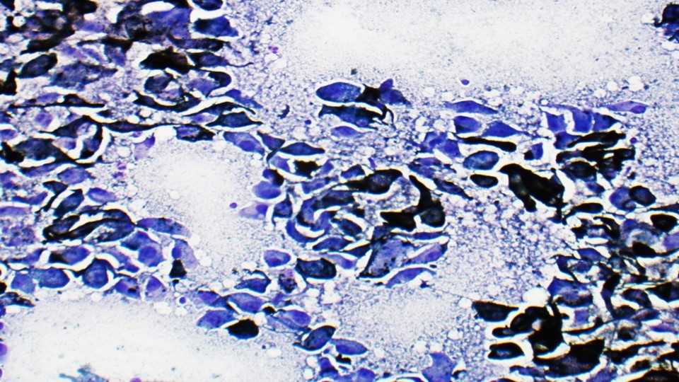
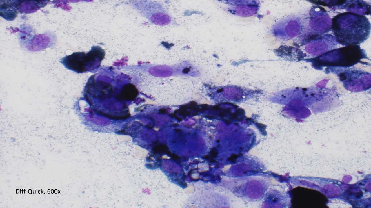
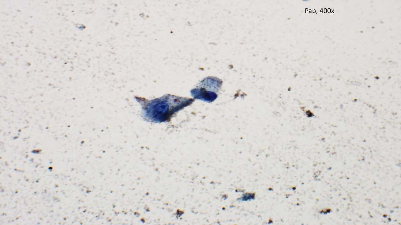
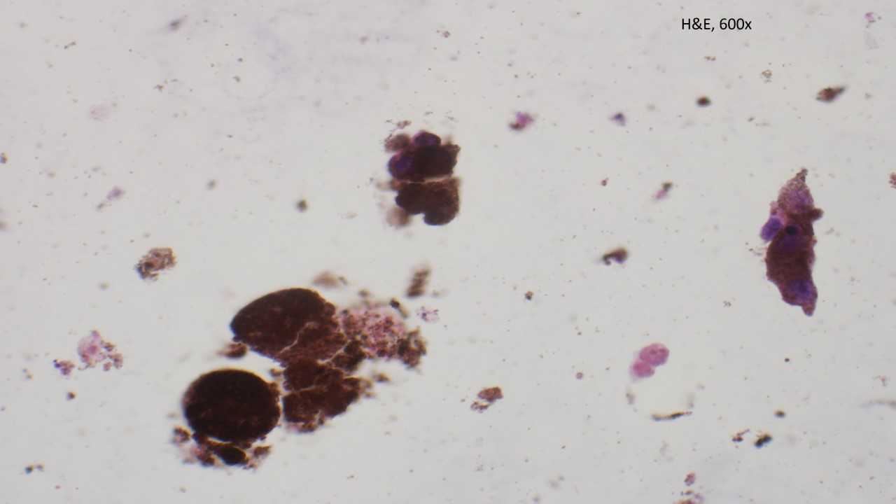
The specimen is cellular and consists of a dyshesive population of large atypical plasmacytoid cells with prominent nucleoli and fine granular cytoplasmic pigment obscuring some of the cellular details.
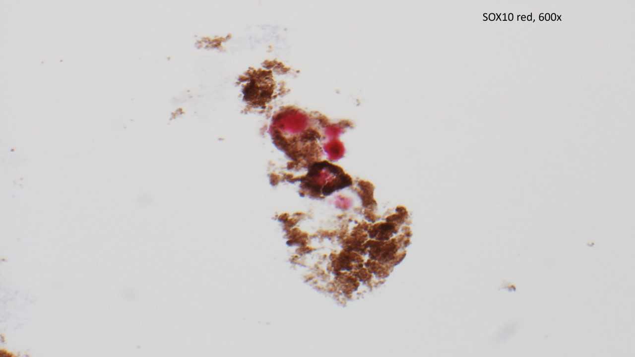
Because the patient had a prior history (invasive melanoma of the forehead), a single immunohistochemical stain was performed on this case. The cells were positive for SOX10, confirming the morphologic impression of metastatic melanoma.
Primary melanomas of the parotid gland are extremely rare, and most cases represent metastasis from head and neck primaries. Metastatic melanoma is the second most common metastatic tumor to the parotid gland (after squamous cell carcinoma).
Case contributed by: Frida Rosenblum, M.D., Associate Professor, Anatomic Pathology, UAB Pathology