Case History
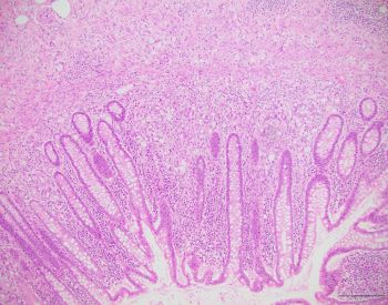
Appendectomy specimen with a clinical diagnosis of acute appendicitis.
What is the diagnosis?
A. Mucinous carcinoid
B. Signet ring cell adenocarcinoma
C. Goblet cell adenocarcinoma
D. Metastatic adenocarcinoma
Answer: C. Goblet cell adenocarcinoma.
Discussion
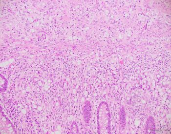
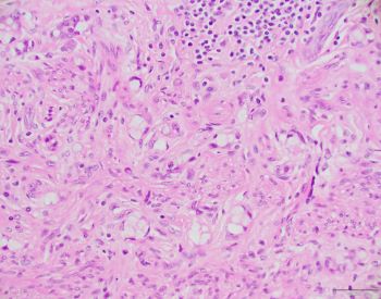
Tumor cells are present in clusters, appearing as goblet/signet ring cells. The overlying crypt epithelium is not involved. The histologic features are diagnostic of this entity, which is characteristically seen in the appendix with no discrete gross lesion. Immunohistochemical studies are not required. These tumors have to be graded (See 2019 WHO classification) based on the poorly cohesive/solid sheets of tumor cells.
Case contributed by: Deepti Dhall, M.D., Professor, Associate Director, Anatomic Pathology, GI Section Head, UAB Pathology