Case History
29 yo P1001 di/di twins at 33 weeks. Pre-eclampsia, selective IUGR baby B, PPROM.
What is the likely diagnosis?
A. Maternal vascular malperfusion
B. Twin-twin transfusion
C. Massive perivilllous fibrinoid deposition
D. Infection
Answer: C. Massive perivillous fibrinoid deposition
Discussion
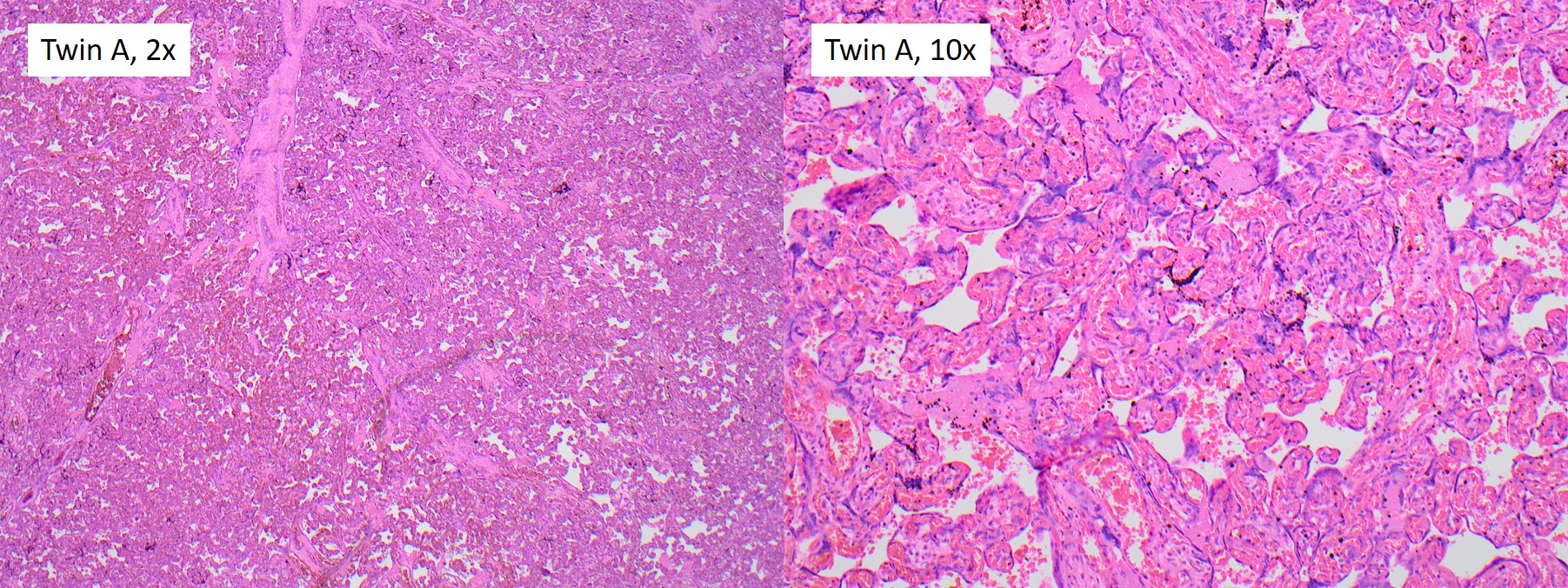
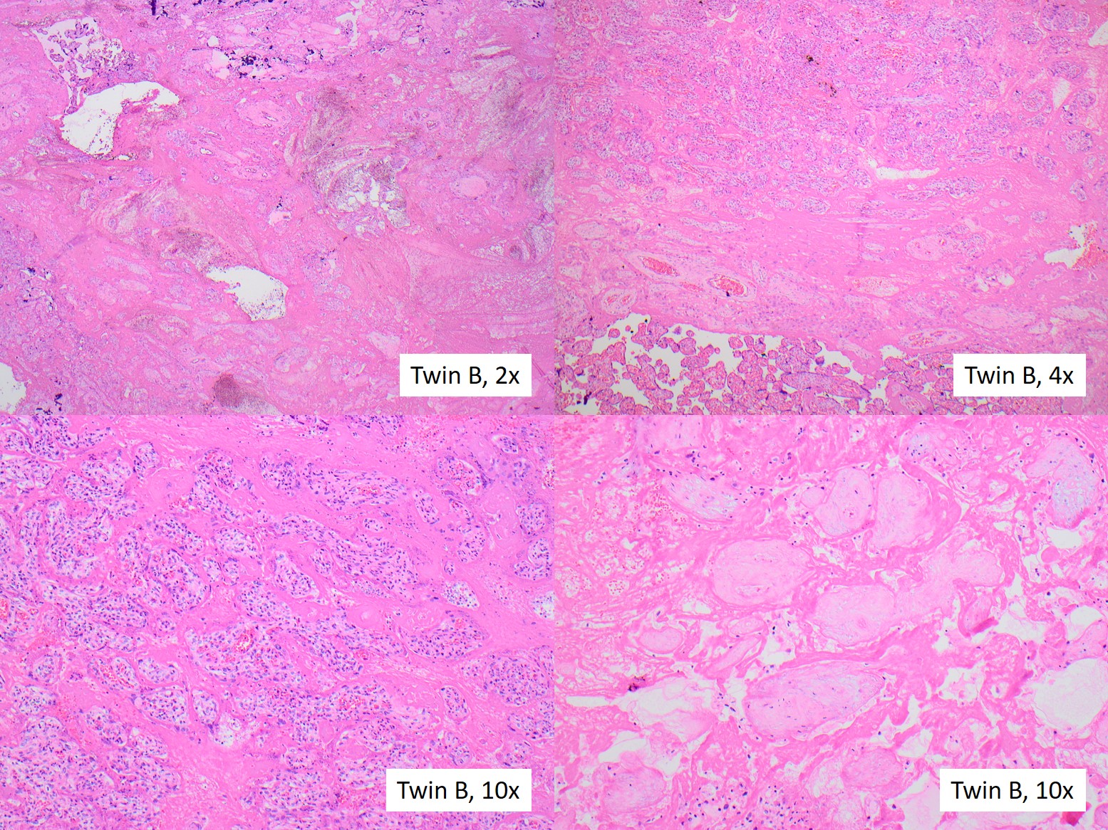
This is a case of massive perivillous fibrinoid deposition (MPVFD), which is discordant in the placental domains of dichorionic twins. The gross photograph shows extensive deposition of firm, tan material entirely confined to the domain of twin B, visible over the near-entirety of the maternal surface with transmural marbling of the majority of the parenchyma. Histology showed extensive regions of chorionic villi cloaked by fibrinoid material involving approximately 70% of the sampled material. Panel A in the photomicrograph shows a low power view of an affected region in the domain of twin B, and dark pink fibrinoid is seen diffusely expanding the intervillous space. Panel B is a higher power view of an affected region (top) adjacent to an unaffected region of twin B domain. Panel C and D are higher power views showing affected parenchyma of varying age with the maternal space completely filled by fibrinoid, with more viable chorionic villi in C and completely involuted chorionic villi in D. Twin A placental domain gross and histologic examination was normal.
MPVFD is a severe placental lesion characterized by increased perivillous fibrinoid expanding the intervillous space, surrounding and separating the chorionic villi. The fibrinoid material may be most prominent in the basal region (aka maternal floor infarction (MFI)), or may be more generalized as seen in this case. Although the gross appearance may mimic infarction, the lesion is not due to chronic ischemia, but rather though to be the result of an alloimmune process (e.g. maternal anti-fetal reaction). MPVFD is highly associated with fetal growth restriction and perinatal morbidity and mortality. MPVFD is a rare entity, and discordant presentation in only one domain of a twin pregnancy is exceedingly rare. However, discordant MPVFD in diamniotic dichorionic twins has been reported in a handful of cases. Since the process is only involving Twin B in this case, MPVFD likely explains the selective intrauterine growth restriction in twin B. MPVFD is an important lesion to recognize because recurrence risk in subsequent pregnancies is high (variously reported as 12-78%). Evidence suggests that more extensive involvement of the placenta is associated with worse outcomes. No standardized system for diagnosis exists at this time, but semiquantitative criteria have been proposed for the diagnosis of MPVFD. One useful schema suggests distribution into the following patterns:
1. Classic: entire maternal surface encased in fibrinoid with >3 mm of thickness on at least one slide
2. Borderline: 25%–50% involvement of villi on at least one slide with near-transmural distribution
3. Transmural: extension of fibrinoid to involve the entire thickness of the parenchyma, involving >50% villi on at least one slide
References:
1. Feist H, Blöcker T, Rau G, Hussein K. Discordancy for Placental Massive Perivillous Fibrin Deposition and Fetal Growth in Dichorionic Twins after In Vitro Fertilization. Pediatr Dev Pathol. 2015 Sep-Oct;18(5):405-9. doi: 10.2350/15-02-1609-CR.1. Epub 2015 Apr 23. PMID: 25905453.
2. Faye-Petersen O, Sauder A, Estrella Y, Heller DS. Dichorionic Twins Discordant for Massive Perivillous Fibrinoid Deposition: Report of a Case and Review of the Literature. Int J Surg Pathol. 2018 Feb;26(1):41-46. doi: 10.1177/1066896917720029. Epub 2017 Jul 9. PMID: 28691603.
3. Romero R et al., Maternal floor infarction/massive perivillous fibrin deposition: a manifestation of maternal antifetal rejection? Am J Reprod Immunol. 2013 Oct;70(4):285-98. PMID: 23905710
4. Faye-Petersen OM and Ernst LM. Maternal Floor Infarction and Massive Perivillous Fibrin Deposition. Surg Pathol Clin. 2013 Mar;6(1):101-14. PMID: 26838705
5. Lampi K, Papadogiannakis N, Sirotkina M, Pettersson K, Ajne G. Massive perivillous fibrin deposition of the placenta and pregnancy outcome: A retrospective observational study. Placenta. 2022 Jan;117:213-218.
Case contributed by: Virginia Duncan, M.D., Assistant Professor, Anatomic Pathology, Perinatal Section Head, UAB Pathology