Case History
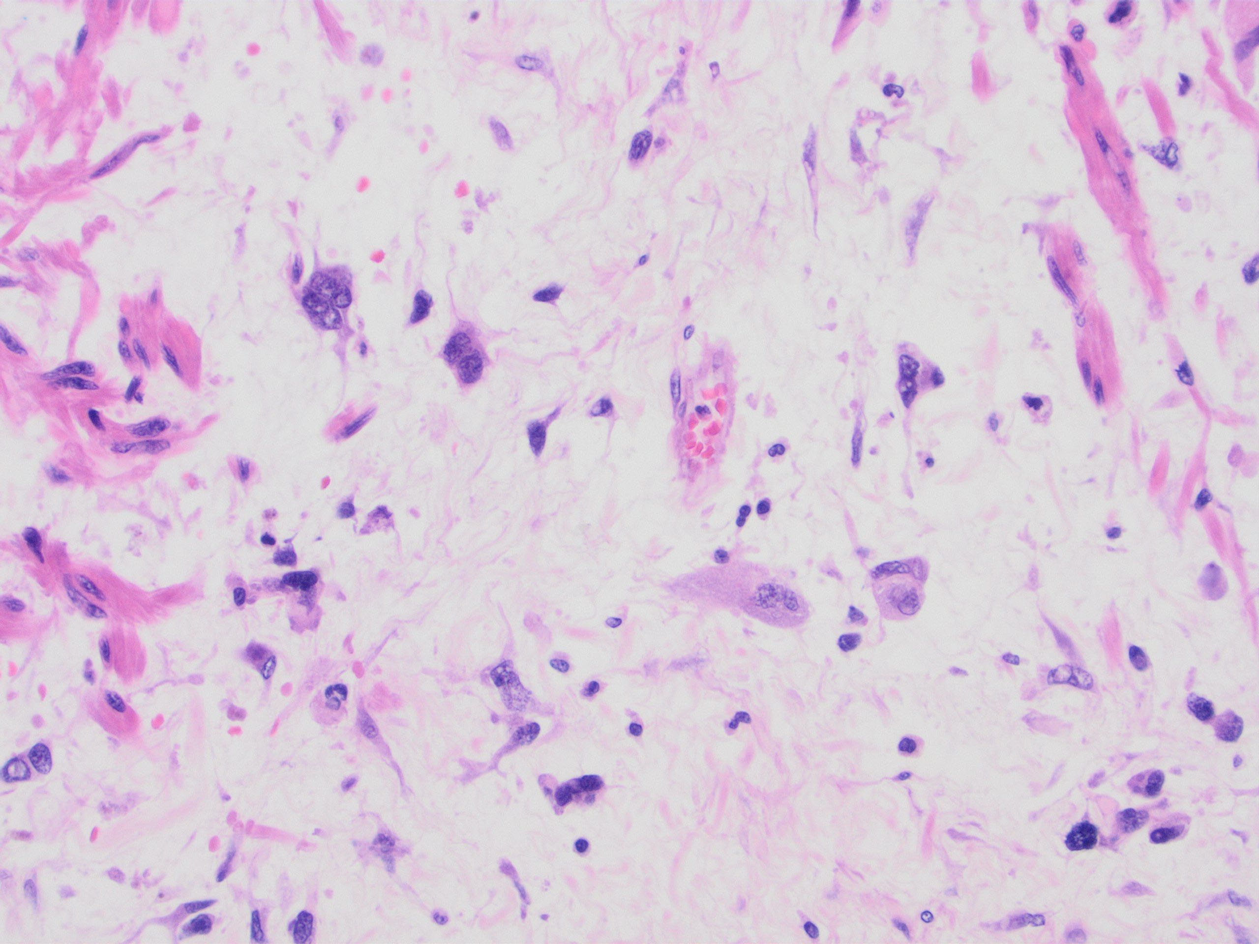
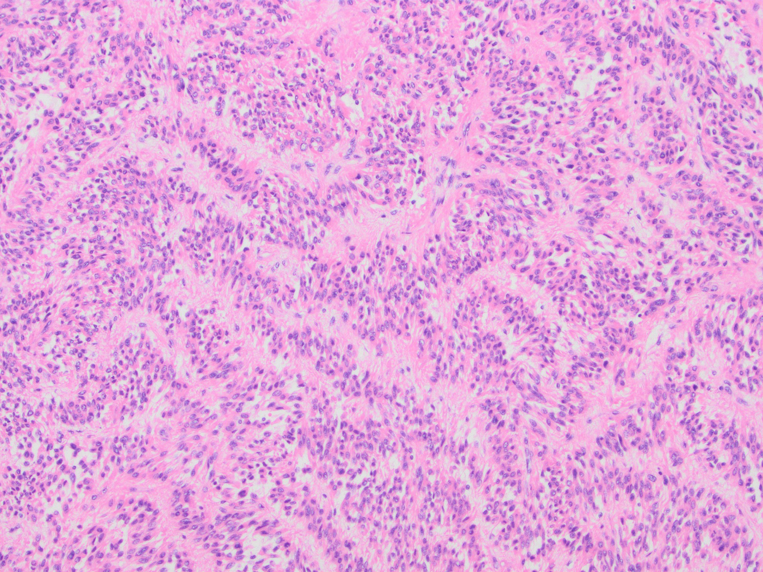
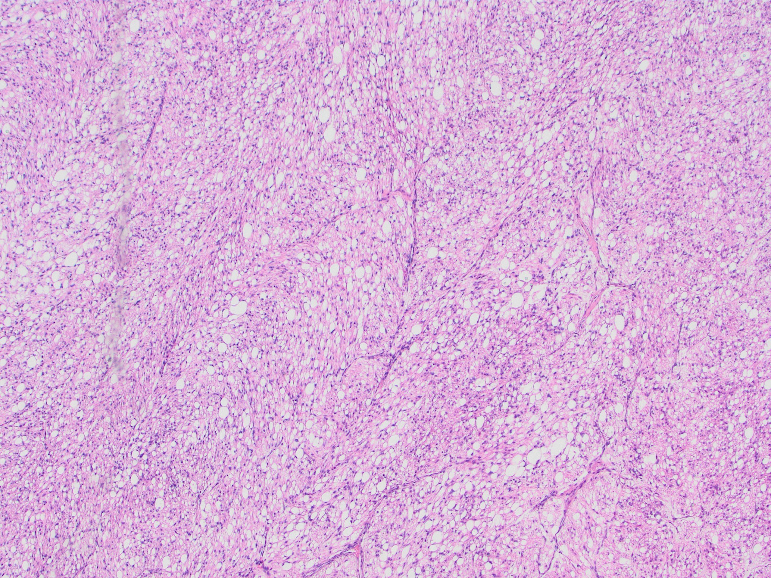
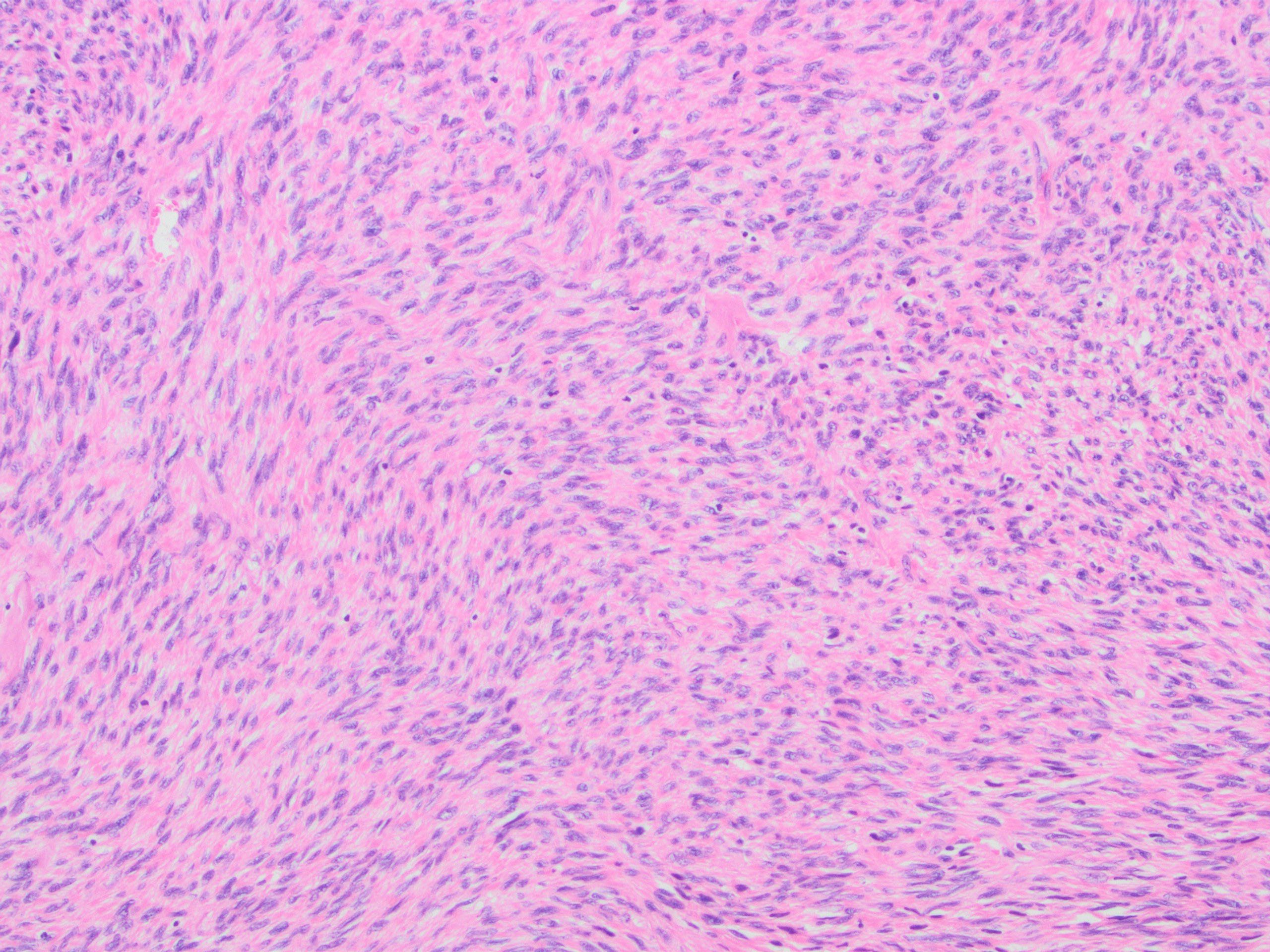
60 yo female with a 28.7 cm solid, hemorrhagic, and degenerated uterine mass. Pelvic soft tissue was involved. The sections show heterogeneous morphology. Mitosis is up to 4 mitosis/10 hpf. Focal tumor necrosis is identified. IHC stains were performed on multiple representative sections that show the tumor is diffusely and strongly positive for desmin, SMM, BCL1, ER, negative for CK, SOX10, S100, SF1, CD10. HMB45 is very focally positive. Solid tumor cancer panel shows no clinically significant sequence variants, fusion genes or copy number alterations. MDM2 FISH testing shows non-amplified.
What is the likely diagnosis?
A. Uterine leiomyosarcoma
B. Endometrial stromal sarcoma
C. PEComa
D. Dedifferentiated liposarcoma
Answer: A. Uterine leiomyosarcoma
Case contributed by: Xiao Huang, M.D., Ph.D., Assistant Professor, Anatomic Pathology, UAB Pathology