Case History
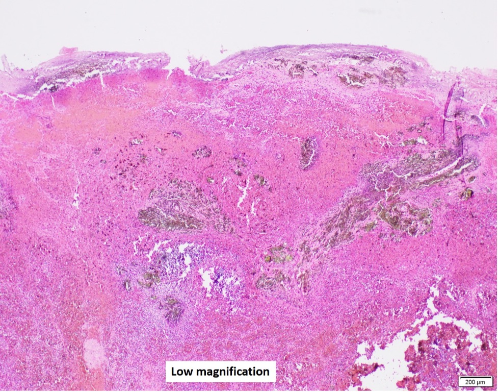
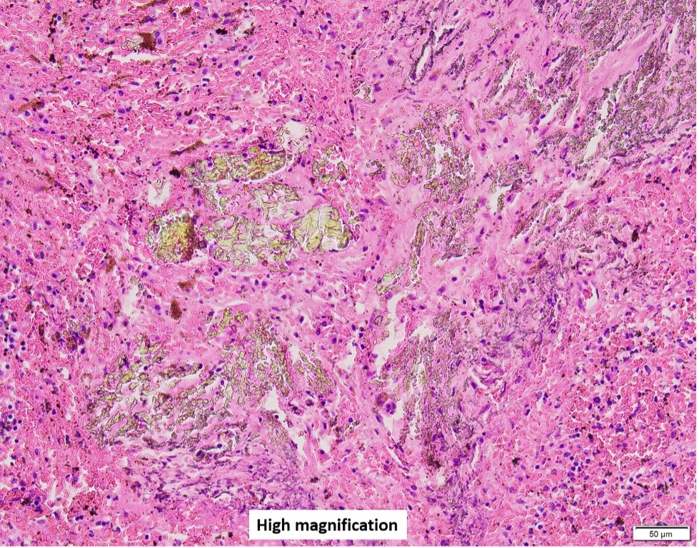
A female in her mid-40s was admitted for splenectomy due to splenomegaly. The resected spleen weighed 655 grams and sections showed multiple tan, plaque-like lesions ranging in size 0.1-0.7 cm. Figures show microscopic findings of the lesion.
Choose the condition(s) associated with the findings.
A. Splenic vein thrombosis
B. Lymphoma
C. Sickle cell disease
D. Portal hypertension
Answer: all of the above
The patient with a history of sickle cell disease required splenectomy due to LLQ pain attributed to splenomegaly.
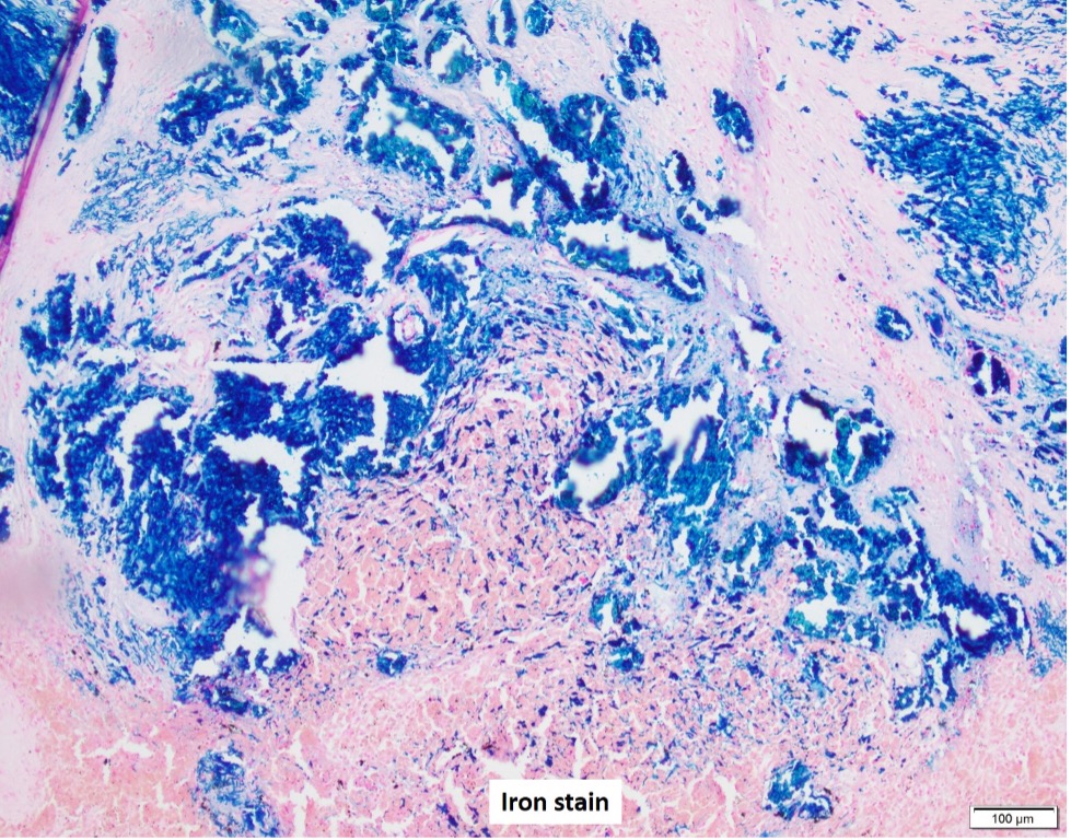
The pictures show Gamna-Gandy bodies, which are foci of organized hemorrhage, appear as yellow-brownish foci grossly, and are associated with a variety of conditions including portal hypertension, sickle cell disease, hemolytic anemia, splenic vein thrombosis, lymphoma, etc (See Ref. 1).
Microscopically the lesions consist of dense fibrosis with refractile, yellow-to-brown-to-black iron and calcium deposition with/without granulomatous reaction (See Figure: iron stain for Gamna-Gandy bodies).
Important differential diagnosis includes infection, which can be achieved by proper special stains.
References:
Piubelli MLM, Clemente LC, Duarte-Neto AN. Gamna-Gandy bodies of the spleen in sickle cell disease. Autops Case Rep. 2019 Mar 22;9(2):e2018076.
Splenic Gandy-Gamna Bodies in American Society of Hematology website.
https://imagebank.hematology.org/imageset/60057/splenic-gandygamna-bodies
Case contributed by: Goo Lee, M.D., Ph.D., Associate Professor, Anatomic Pathology, UAB Pathology