Case History
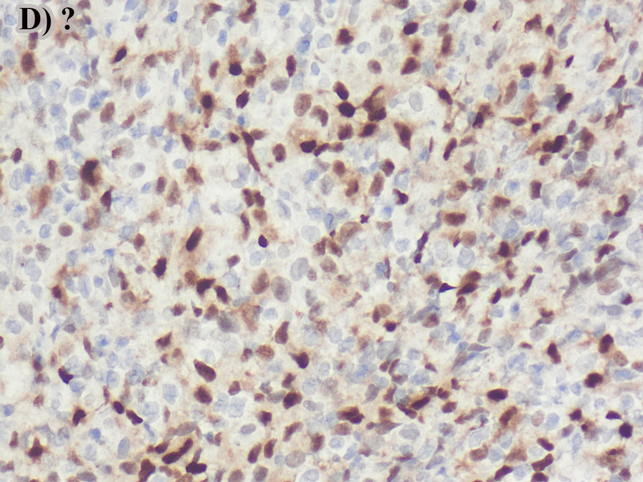
A 60 year-old-male presented with a lytic lesion of the C3 spinal vertebra. What is the diagnosis? Identify the immunostain in Figure D.
What is the best diagnosis?
A. Reactive histiocytic proliferation; cyclin D1
B. Langerhans cell Histiocytosis; BRAF V600E
C. Langerhans cell Histiocytosis; cyclin D1
D. Indeterminate cell Histiocytosis; cyclin D1
Answer: C. Langerhans cell Histiocytosis; cyclin D1
Discussion:
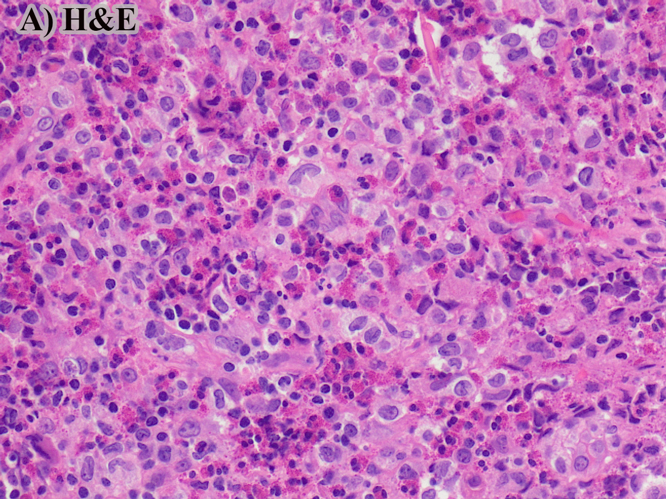
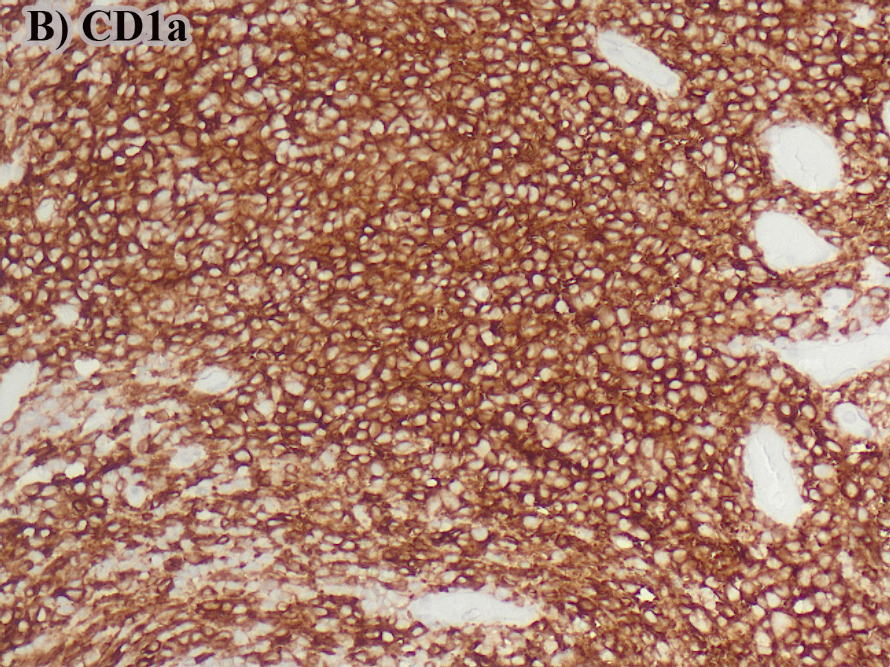
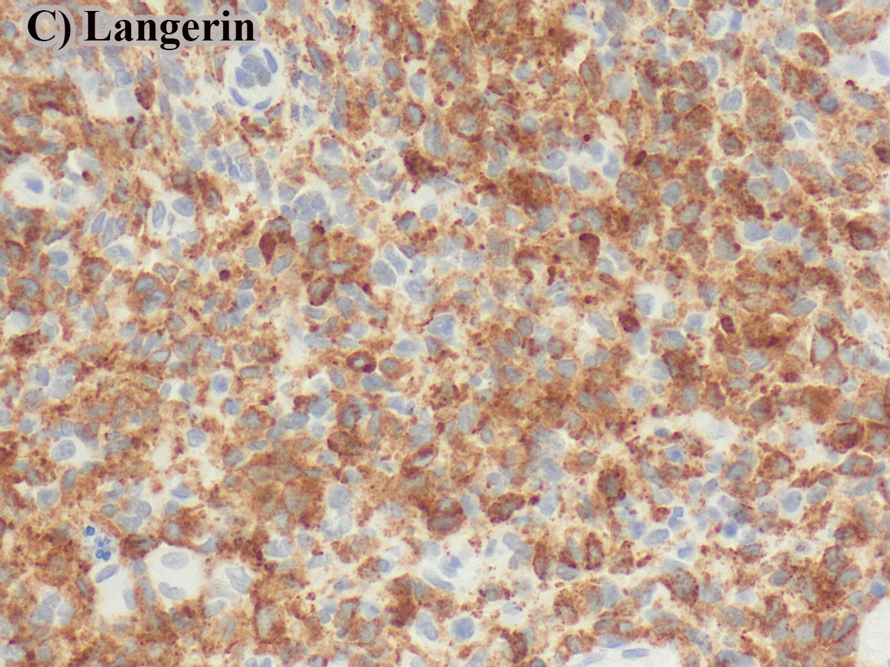
This is a case of Langerhans cells Histiocytosis (LCH) that is characterized by grooved nuclei and abundant pale eosinophilic cytoplasm, admixed with numerous eosinophils. The LCH cells are diffusely positive for CD1a, langerin, and cyclin D1 (strong nuclear and weak cytoplasmic positivity; part D). CyclinD1 expression is usually diffusely positive in neoplastic Langerhans cells, and essentially negative in reactive histiocytic proliferations (PMID: 30124506; PMID: 33177341). This is a consequence of MAPK pathway activation in LCH cases.
BRAF V600E is positive in 50-60% of LCH cases, and the immunostain shows cytoplasmic positivity and absent nuclear staining. CD68 shows cytoplasmic staining in the Langerhans cells and absent nuclear staining. Indeterminate cell Histiocytosis (also known as indeterminate dendritic cell tumor) offers similar morphologic features to LCH but on immunohistochemistry, the indeterminate cells are positive for CD1a and negative for langerin.
Case contributed by: Aishwarya Ravindran, M.D., Assistant Professor, Laboratory Medicine, UAB Pathology