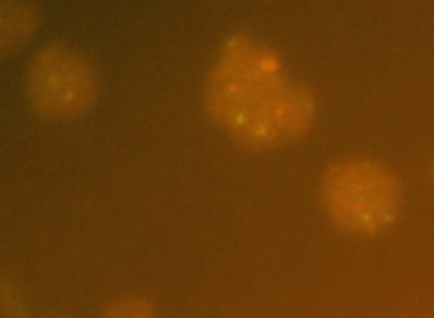
Case history: The patient is a 62 year old female with right kidney mass, who underwent partial nephrectomy. FISH image with TFE3 break apart probe.
What is the diagnosis?
- Clear cell papillary renal cell carcinoma.
- Papillary renal cell carcinoma.
- Clear cell renal cell carcinoma.
- Metastatic carcinoma from ovary.
- MiT family translocation renal cell carcinoma
Answer: “E”, MiT family translocation renal cell carcinoma
Discussion
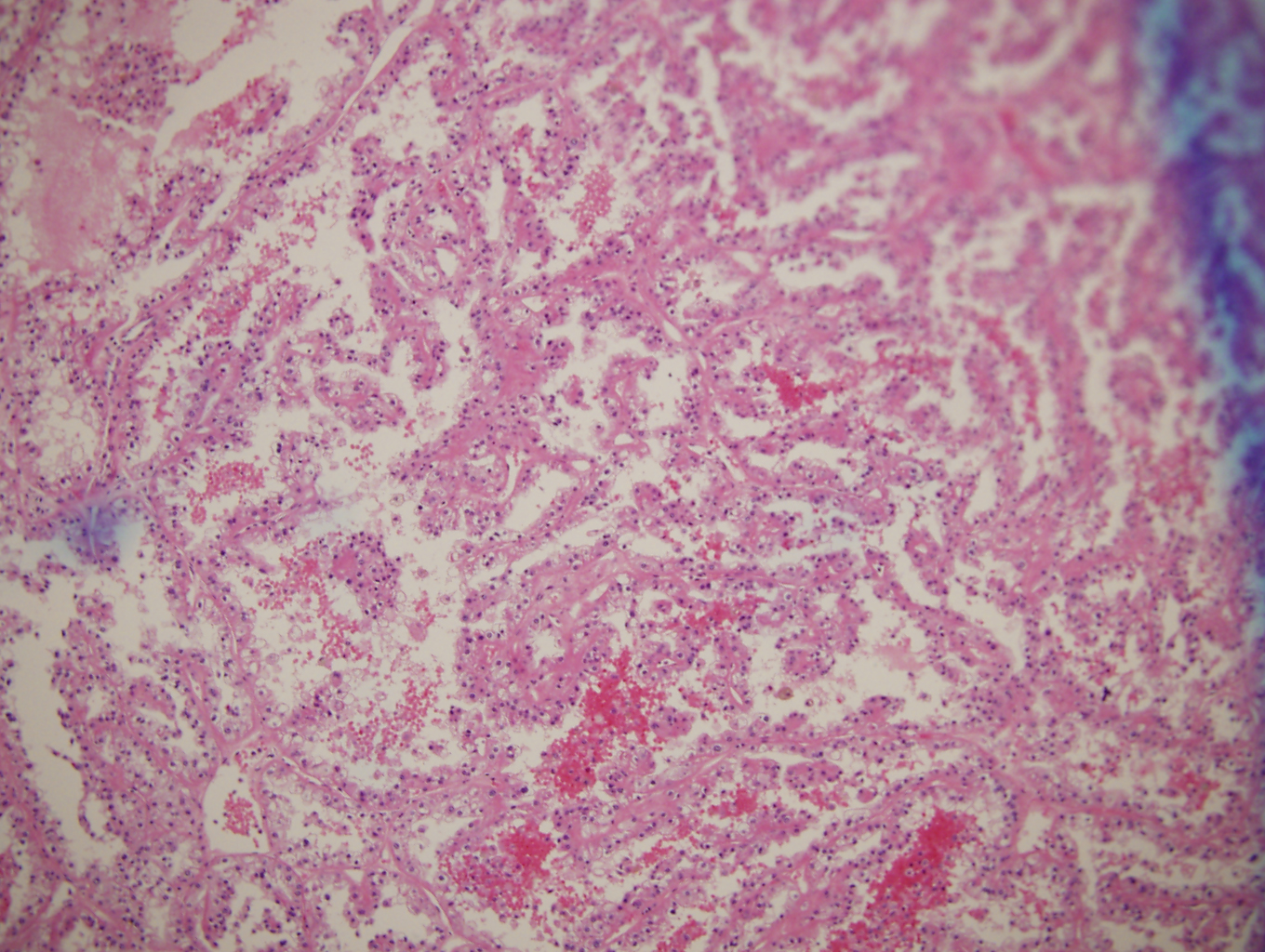
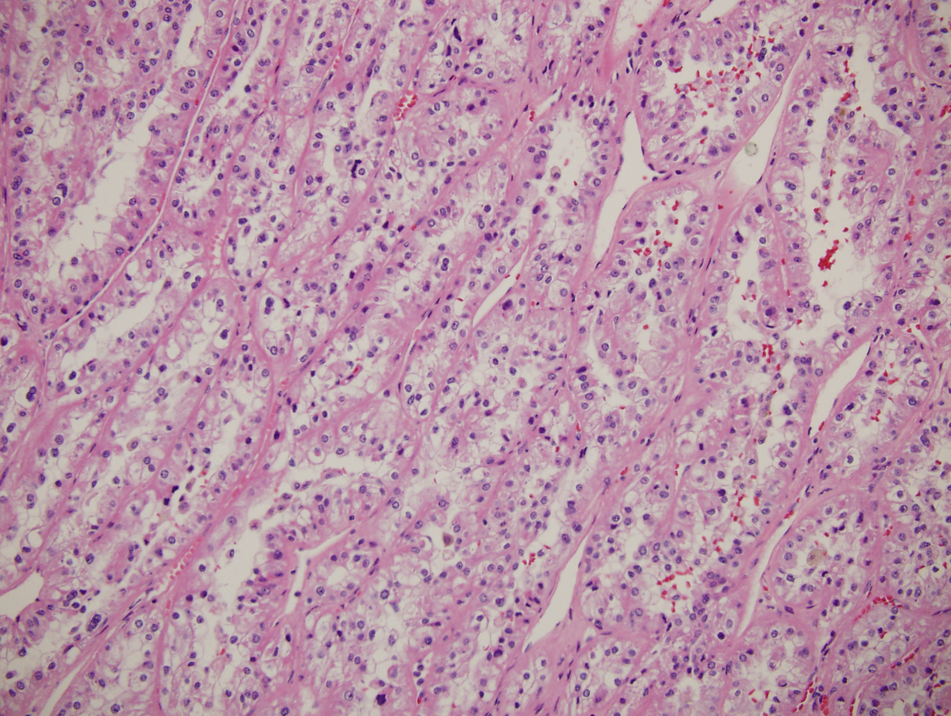
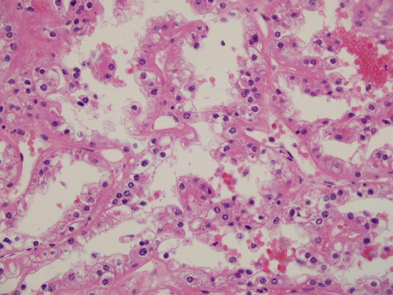
The sections show papillary and solid alveolar growth pattern, composed of clear to eosinophilic cells with voluminous cytoplasm. Tumor cells are positive for Cathepsin K, PAX8 and negative for CK7. Fluorescent in situ hybridization (FISH) with TFE3 break apart probe shows split signals, confirming TFE3 rearrangement on chromosome Xp11. TFE3 is a member of the MiT family of transcription factors. MiT Family Translocation carcinoma is more common in children and young adults, however, it can occur in any age.
Case contributed by Shuko Harada, M.D., Associate Professor, Anatomic Pathology, UAB Department of Pathology