Case history
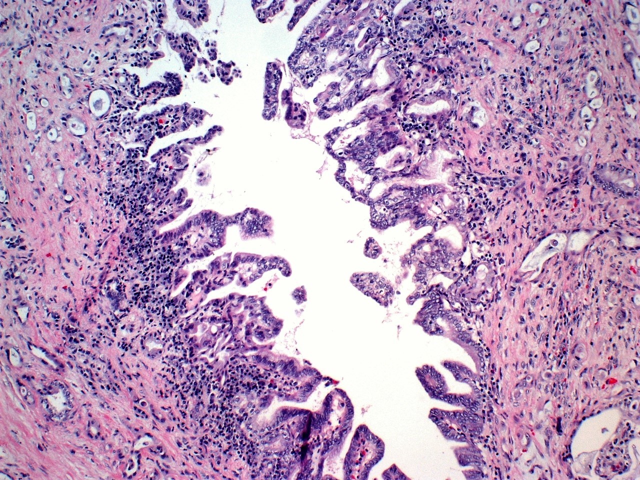
A pancreatic head mass was discovered in a 50-year-old man with chronic pancreatitis. Grossly, the lesion was ill-defined and notable for obstructing the main pancreatic duct while the common bile duct and ampulla were probe patent. The patient noted a mother who died of pancreatic cancer at 55 and that he’s been hospitalized for pancreatitis several times since his 20s. What is the underlying tumor and which of the following genes is not associated with hereditary pancreatitis?
- Pancreatic ductal adenocarcinoma, Mothers against decapentaplegic homolog 4 (SMAD4)
- Cholangiocarcinoma, Cystic fibrosis transmembrane conductance regulator (CFTR) mutations
- Acinar cell carcinoma, Serine protease inhibitor Kazal type 1 (SPINK1) mutations
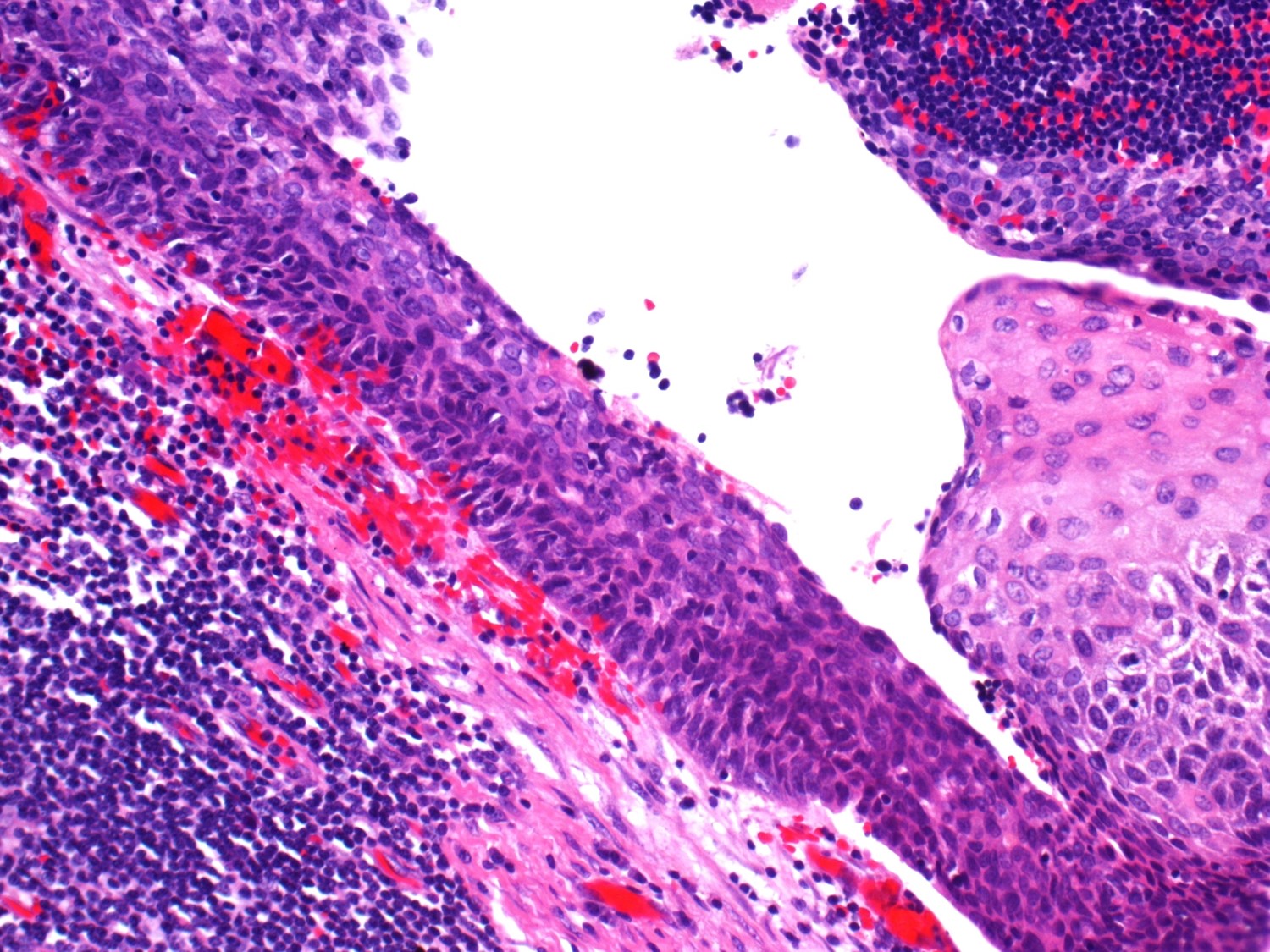
A 5-cm peripancreatic mass was noted on abdominal CT in a 60 year old man during evaluation post motor vehicle accident. EUS-FNA of the mass was notable for acellular keratinous debris. Resection was performed. Grossly, the lesion was unilocular with thick caseous cystic contents. What is the lesion?
- Mucinous cystic neoplasm (mucinous cystadenoma)
- Serous cystadenoma
- Lymphoepithelial cyst
- Intraductal papillary mucinous neoplasm
Answers:
1: “A” Pancreatic ductal adenocarcinoma, Mothers against decapentaplegic homolog 4 (SMAD4)
2: “C” Lymphoepithelial cyst
Case contributed by Leona Council, M.D., Assistant Professor, Anatomic Pathology, UAB Department of Pathology