Case history
71-year-old male veteran presenting with hematuria. Two ulcerated lesions identified upon gross examination. The diagnosis is:
- Plasmacytoid variant of urothelial carcinoma
- Plasmacytoma
- Melanoma
- Metastatic signet ring cell carcinoma
Answer: A. Plasmacytoid variant of urothelial carcinoma
Brief explanation of the answer:
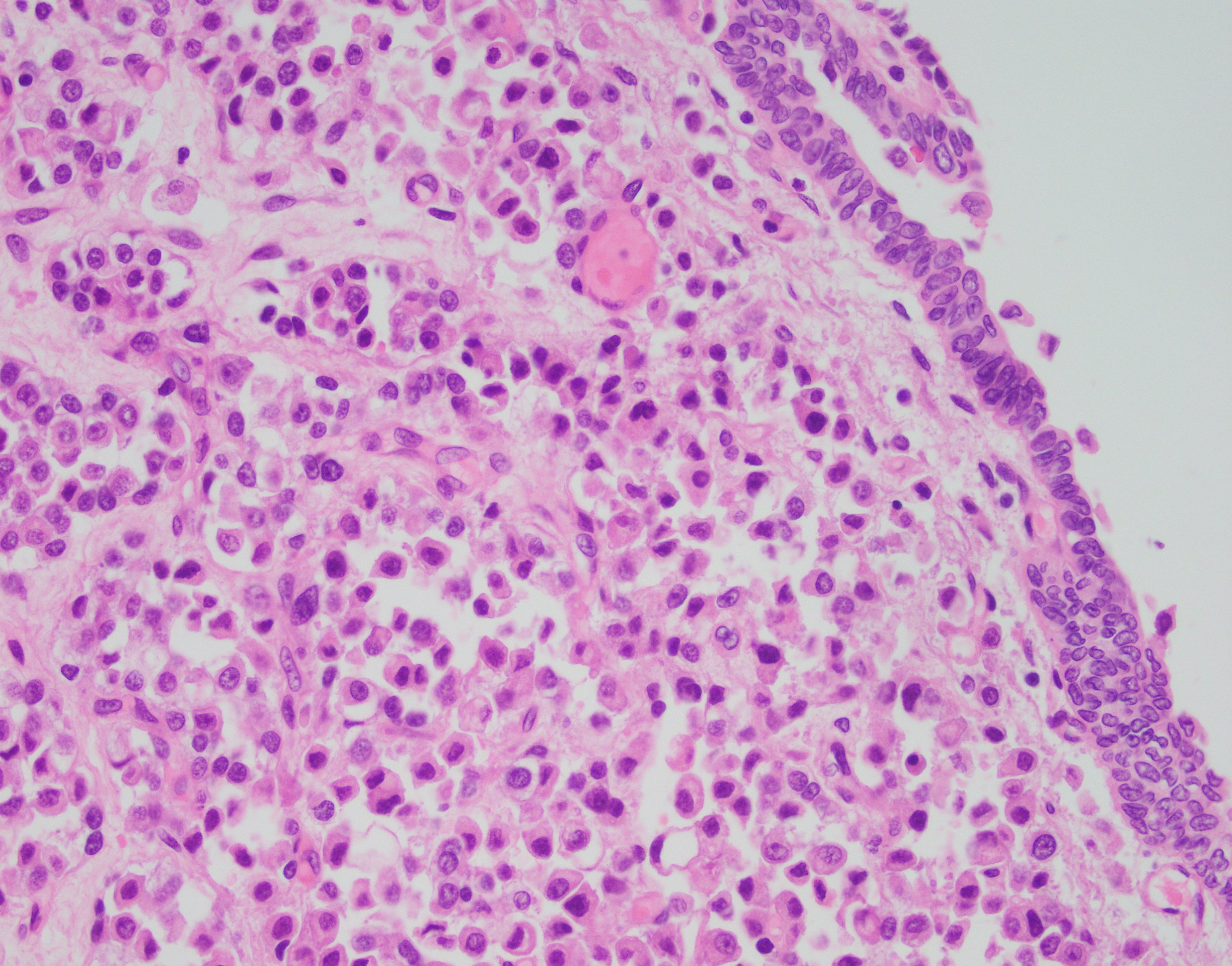
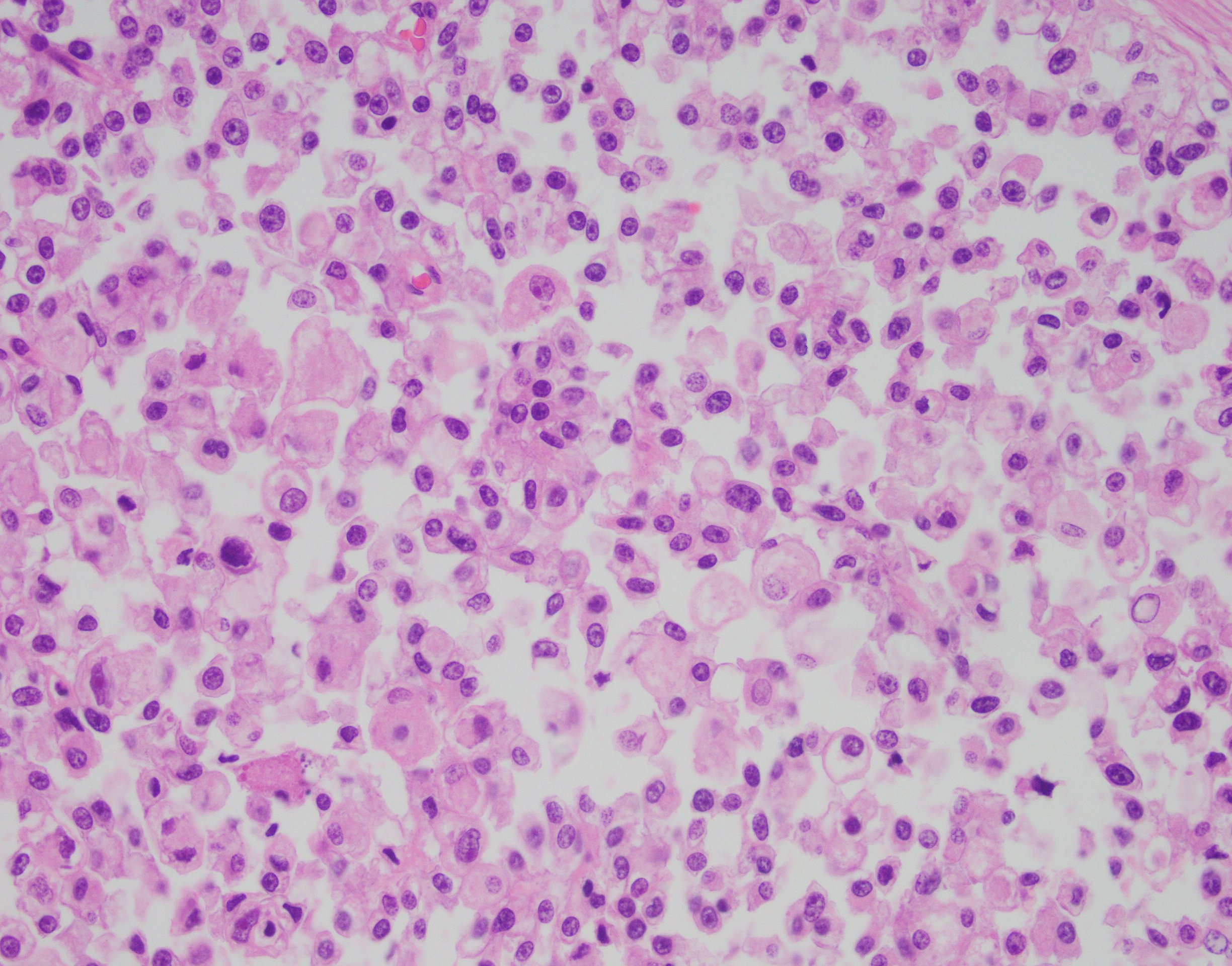
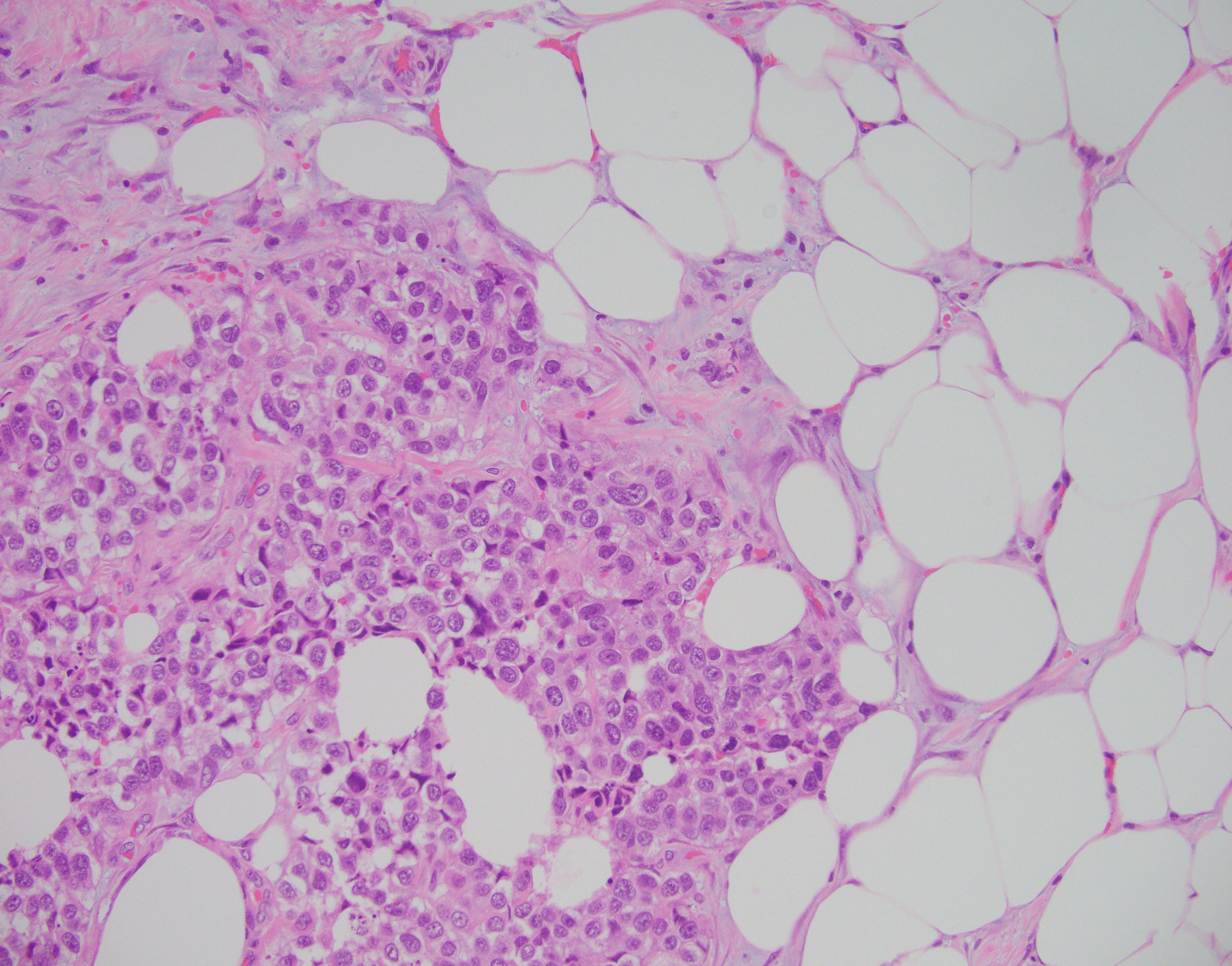
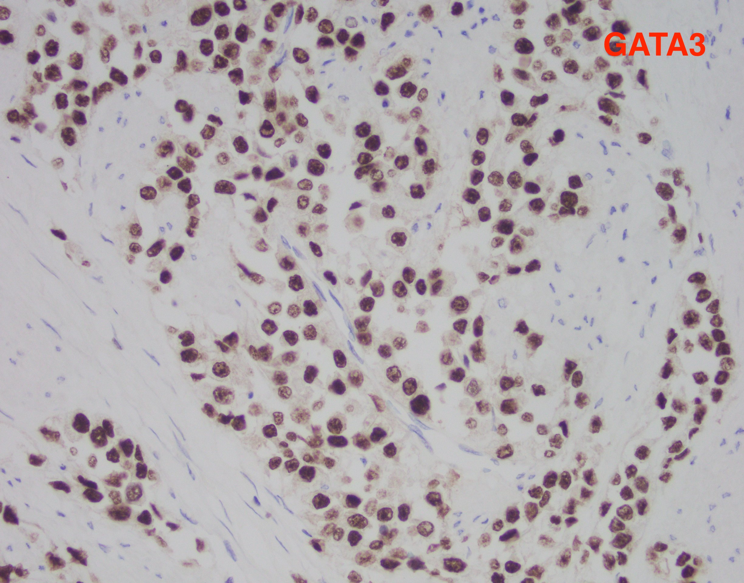
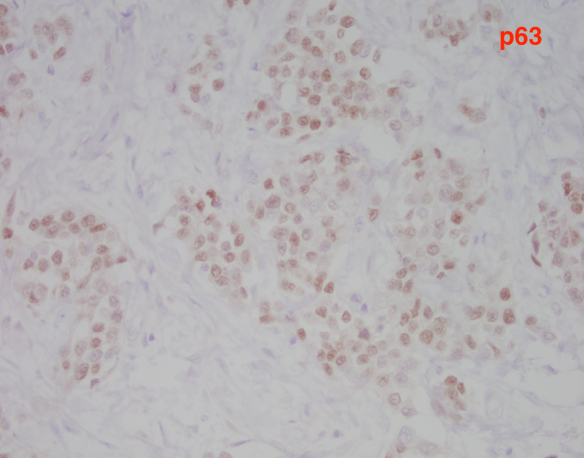
The findings in this case show a diffuse, infiltrative, and discohesive tumor extending into the perivesical adipose tissue. There is extensive perineural and lymphovascular invasion. Focal areas show urothelial carcinoma in situ. Tumor cells have eosinophilic cytoplasm, and nuclei are eccentrically located with focal signet ring cell features. Staining with p63 and GATA3 was performed and shows positivity in both stains.
These features are all consistent with the plasmacytoid variant of urothelial carcinoma. Typically, this variant grows in a linitis plastica-like manner. Tumor infiltration is diffuse, in single cells or cords. Characteristically, tumor cells exhibit eccentric nuclei and/or signet ring cell morphology. Of note, these tumors stain similar to usual urothelial carcinomas, but also strongly express CD138 and lose E-cadherin expression. Most tumors are locally advanced at the time of diagnosis and are highly aggressive.
References:
McKenney, Jesse K. “Overview of Invasive Carcinoma Subtypes”. ExpertPath. https://app.expertpath.com/document/overview-of-invasive-carcinoma-sub-/7ec907d0-a31d-45e6-aef5-3c3aeee18078. Accessed 11/20/2018.
Eble, John N., Grignon, David J., Young, Robert H. “Tumors of the Urinary Tract”. Diagnostic Histopathology of Tumors. Chapter 12: 559-657. 4th edition. 2013.
Case contributed by Joseph Drwiega, M.D., UAB Department of Pathology