Case History
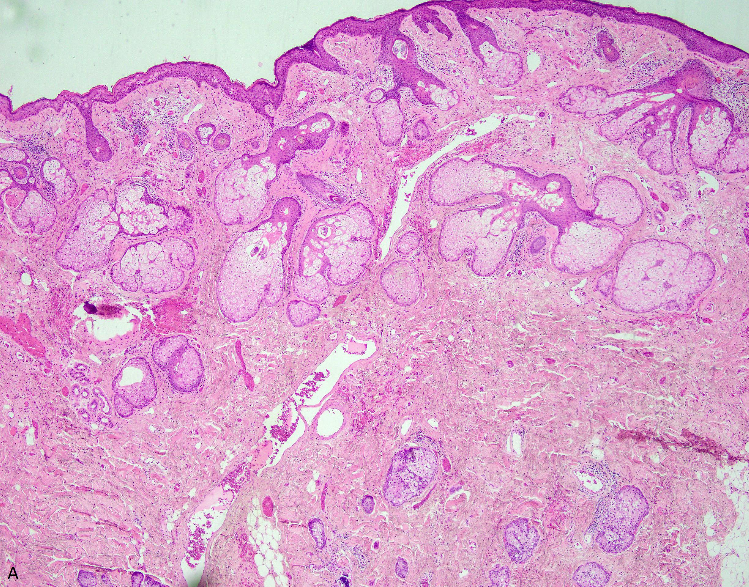
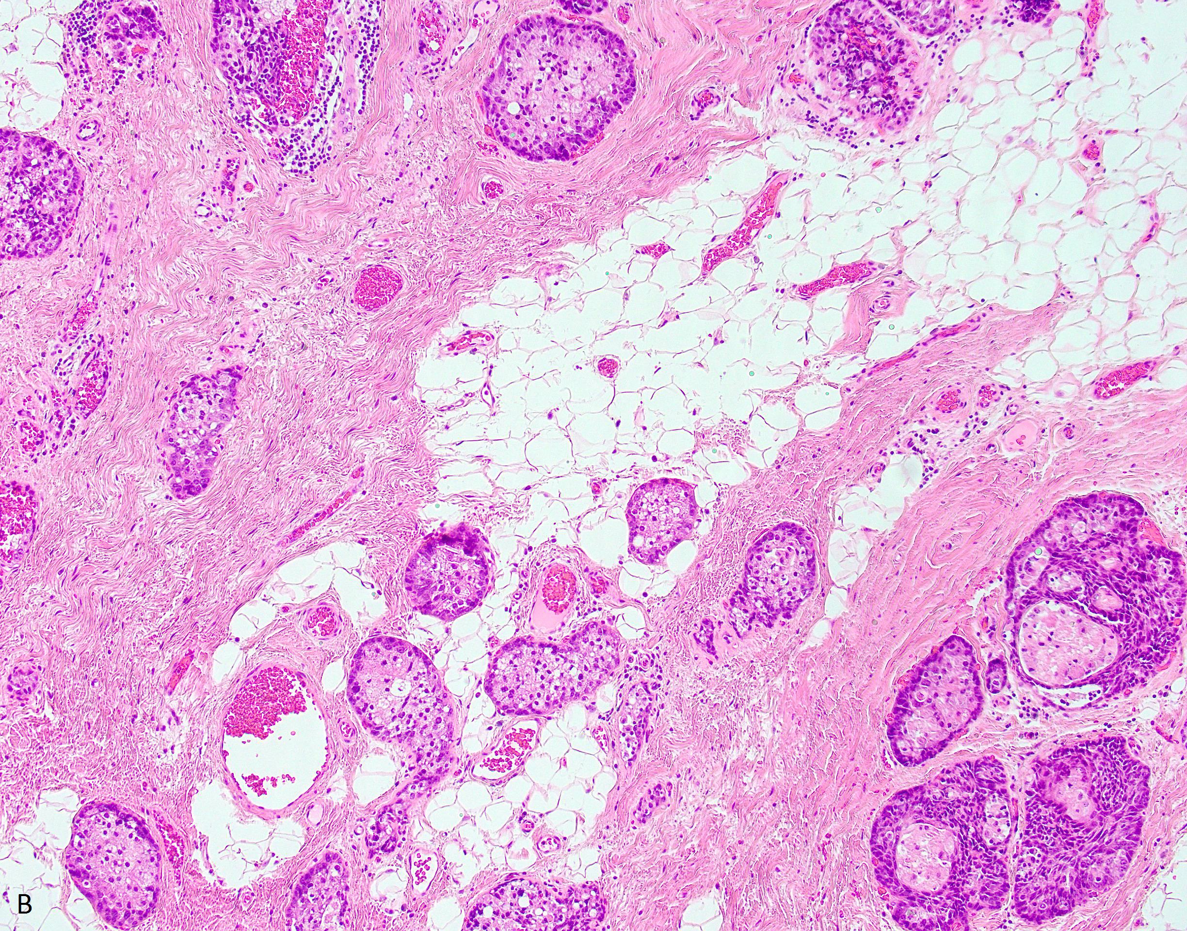
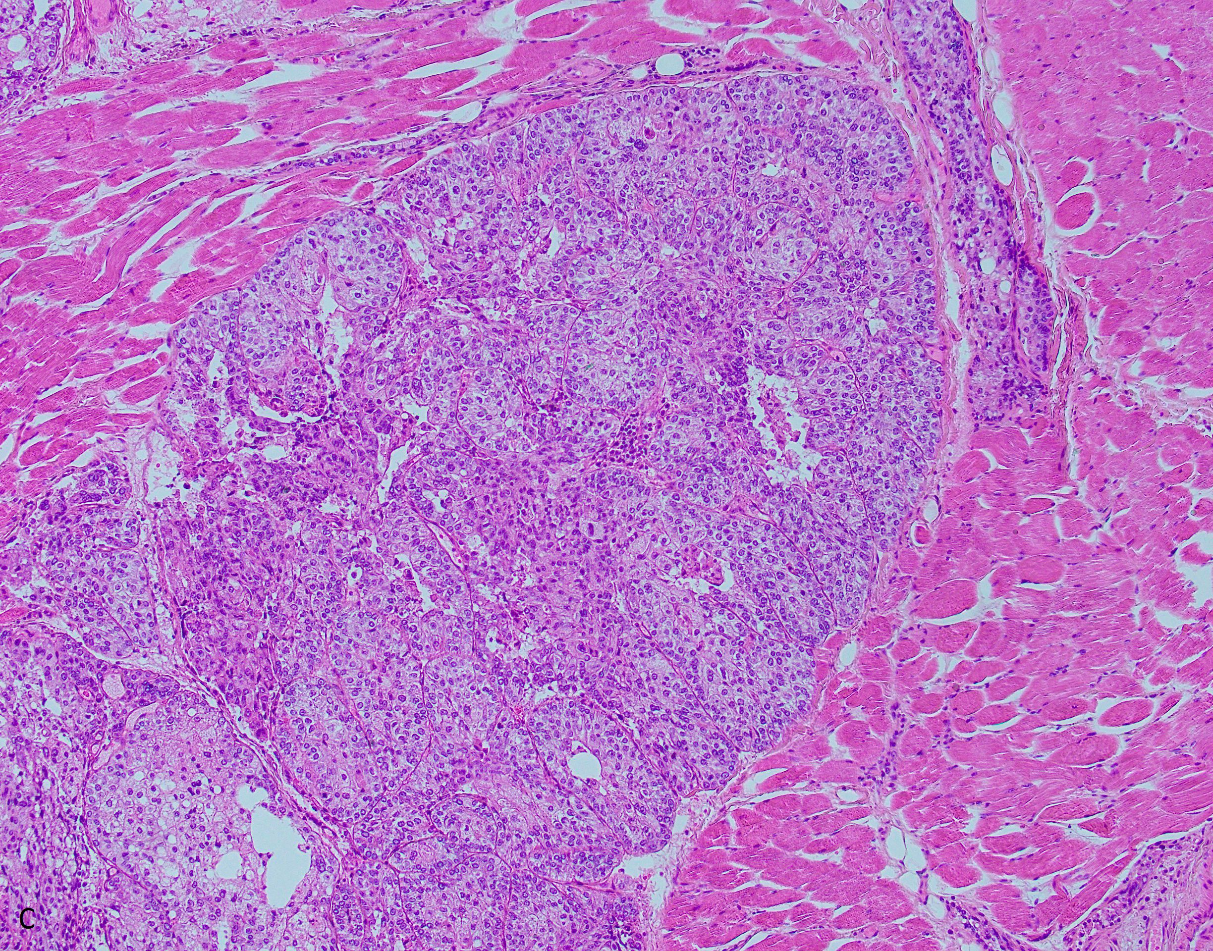
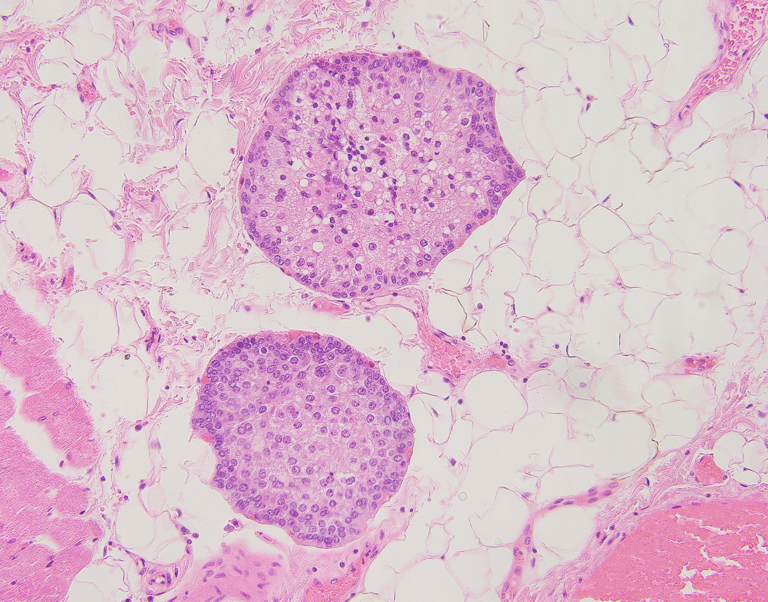
A 62-year-old- male with a history of multiple skin cancers presents to an ENT for a newly discovered firm mass adjacent to his right nasal ala. After biopsy, the mass is resected (Fig A-D).
This tumor is most likely to stain positive for:
- CEA
- Adipophilin
- S100
- Ber-EP4
Answer: “B.”
Brief explanation:
A neoplasm can be seen arising in the dermis exhibiting well circumscribed nests of basaloid cells showing peripheral palisading, focal sebaceous differentiation, moderate nuclear atypia, and increased mitotic activity. Along with adipophilin positivity, these tumors typically show positive immunostaining with keratin, EMA, LeuM1, and androgen receptor. This immunoprofile, along with the histologic features of the tumor, leads to the diagnosis of sebaceous carcinoma. Sebaceous carcinoma often affects areas surrounding the eye, including the eyelid, caruncle, and orbit, but can be found elsewhere in the skin, though the lesions surrounding the eye often exhibit more aggressive behavior. The tumor has been seen in association with Muir-Torre syndrome while other cases have been shown to arise from nevus sebaceous of Jadassohn as well as areas that have been previously irradiated. The differential diagnosis includes basal cell carcinoma with sebaceous differentiation and squamous cell carcinoma with hydropic change.
Case contributed by David Marbury, M.D., UAB Department of Pathology