Case History
A 62 year-old man presenting with jaundice, pancreatic duct/common bile duct dilation and ampullary mass.
What is the most appropriate diagnosis?
- Nondiagnostic specimen
- Negative for malignant cells
- Atypical cells present
- Neoplastic cells present
- Adenocarcinoma
Answer:
D. Neoplastic cells present
Brief explanation of the answer:
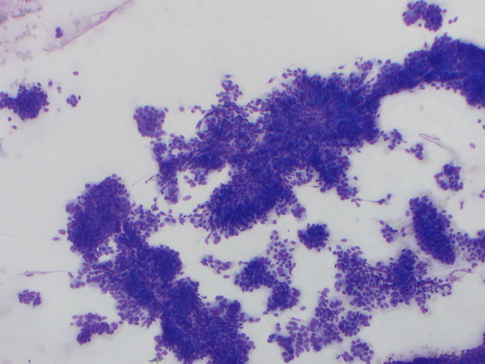
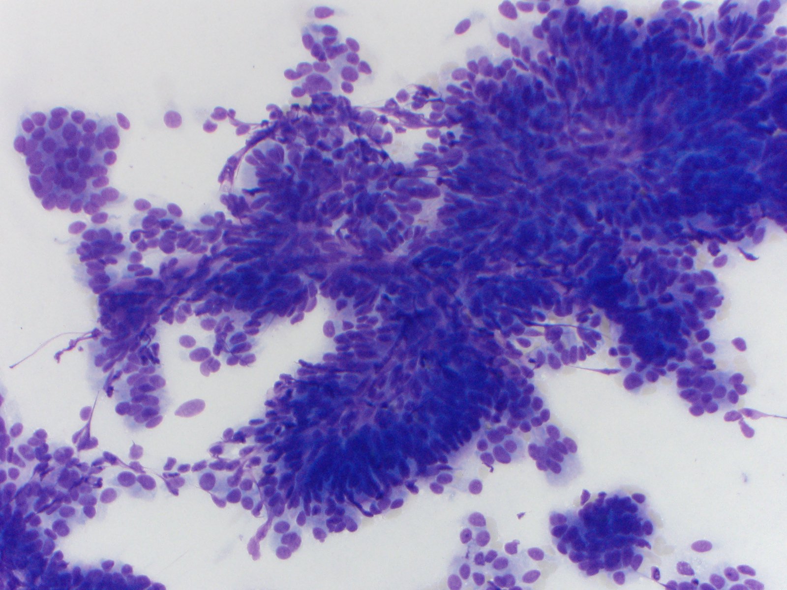
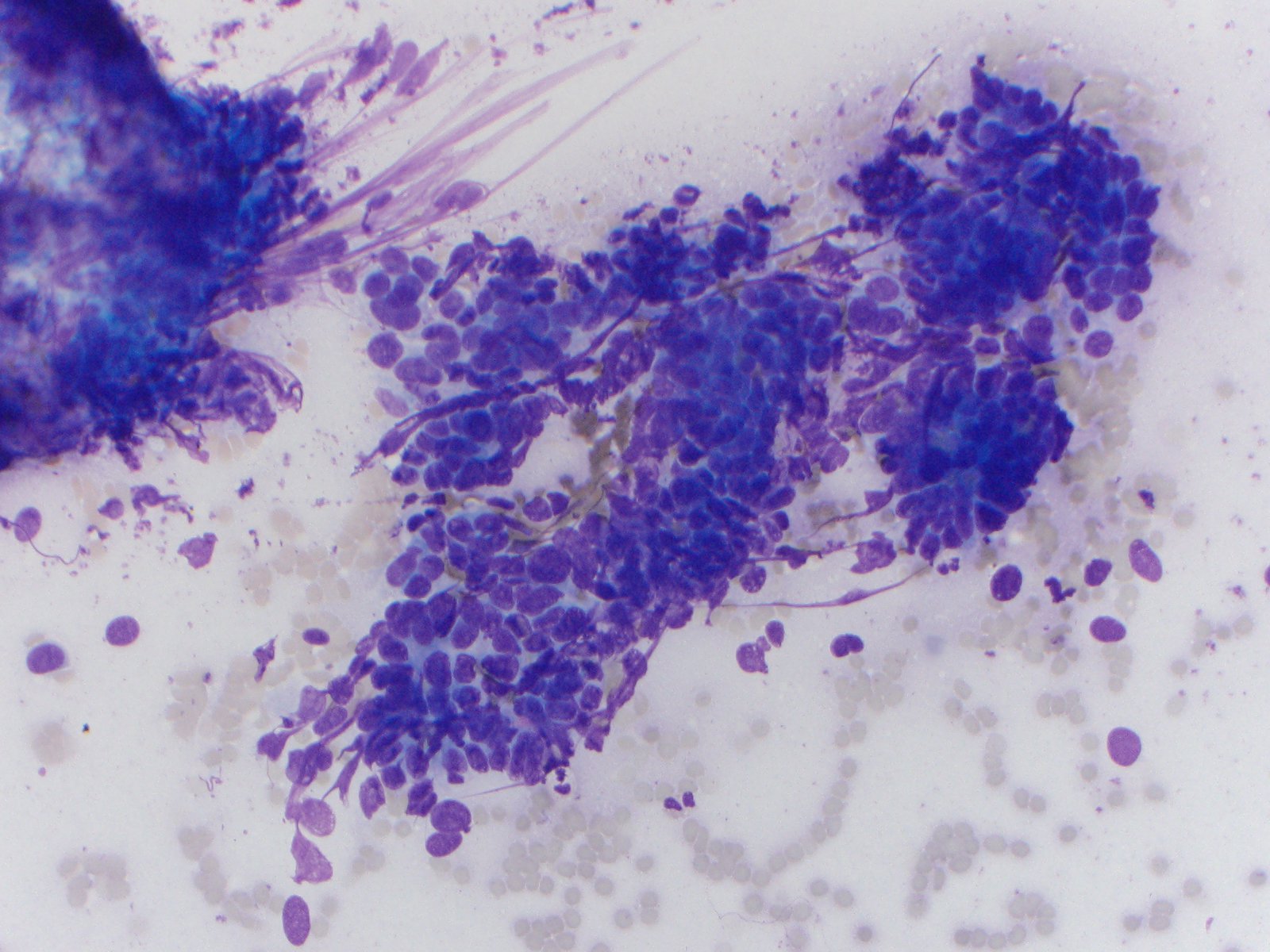
The specimen is highly cellular and consists of abundant fragments of disordered and pseudostratified epithelium with some arborizing and papillary architecture (Figure 1). The cells are monotonous and have oval nuclei, smooth nuclear membranes, no distinct nucleoli and minimal to moderate amounts of cytoplasm (Figure 2). Occasional larger and more pleomorphic cells are noted (Figure 3). No necrosis or single atypical cells are noted in the background.
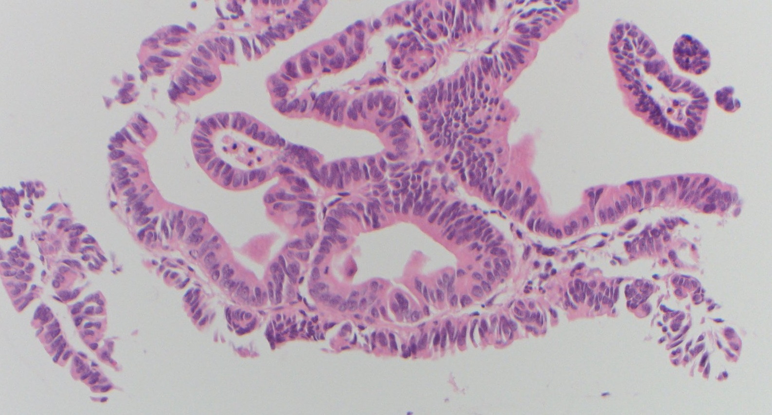
The characteristic features of pancreatic adenocarcinoma including necrotic background, macronucleoli and marked nuclear pleomorphism (>= 4 fold size difference of the nuclei) are not present in this case. In addition, there is intervening stroma in between the glands (Figure 4) and the location (ampulla) of the mass should make us think about the possibility of diagnosis of adenoma. Therefore, these findings are most compatible with an ampullary adenoma with focal high grade dysplasia.
Adenomas are the most common benign lesions of the ampulla. Clinical presentations result from consequences of a mass-effect of the adenoma compressing and impeding biliary or pancreatic outflow. Of note, ampullary adenomas have the potential to undergo malignant transformation to ampullary carcinomas. The diagnosis is usually established on histopathology obtained by duodenoscopy and biopsy. However, only resection can definitively exclude a focus of malignancy.
References:
- Cytology, 4th Edition Diagnostic Principles and Clinical Correlates, by Edmund S. Cibas, MD and Barbara S. Ducatman, MD
- UpToDate: Ampullary adenomas: Clinical manifestations and diagnosis
Case contributed by Yiqin Zuo, M.D., Cytopathology Fellow, Anatomic Pathology, UAB Department of Pathology