Case History
61-year-old female with vaginal wall mass.
Answer choices:
- Fibroepithelial polyp
- Superficial myofibroblastoma
- Keloid
- Cellular angiofibroma

Answer:
B. Superficial myofibroblastoma
Brief Explanation of the answer:
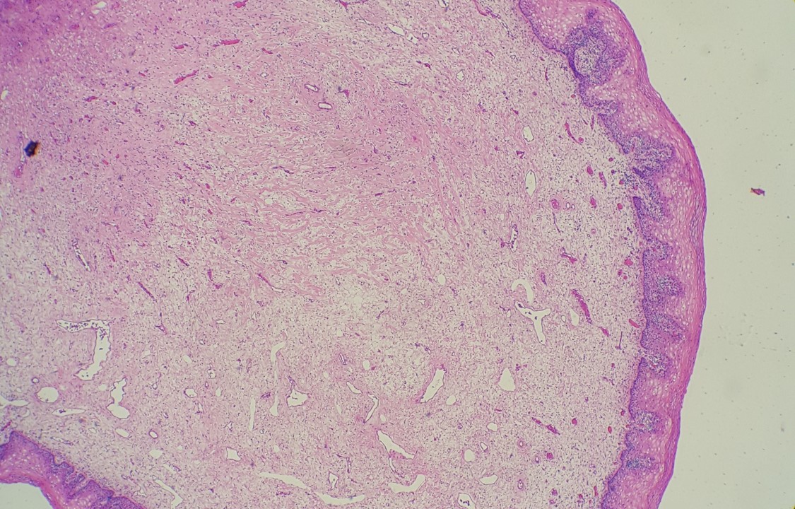
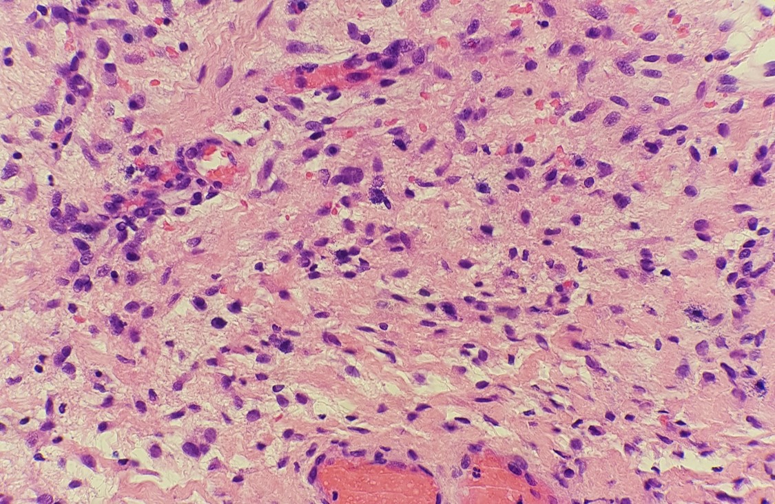
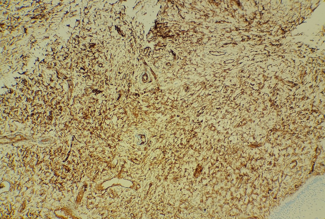
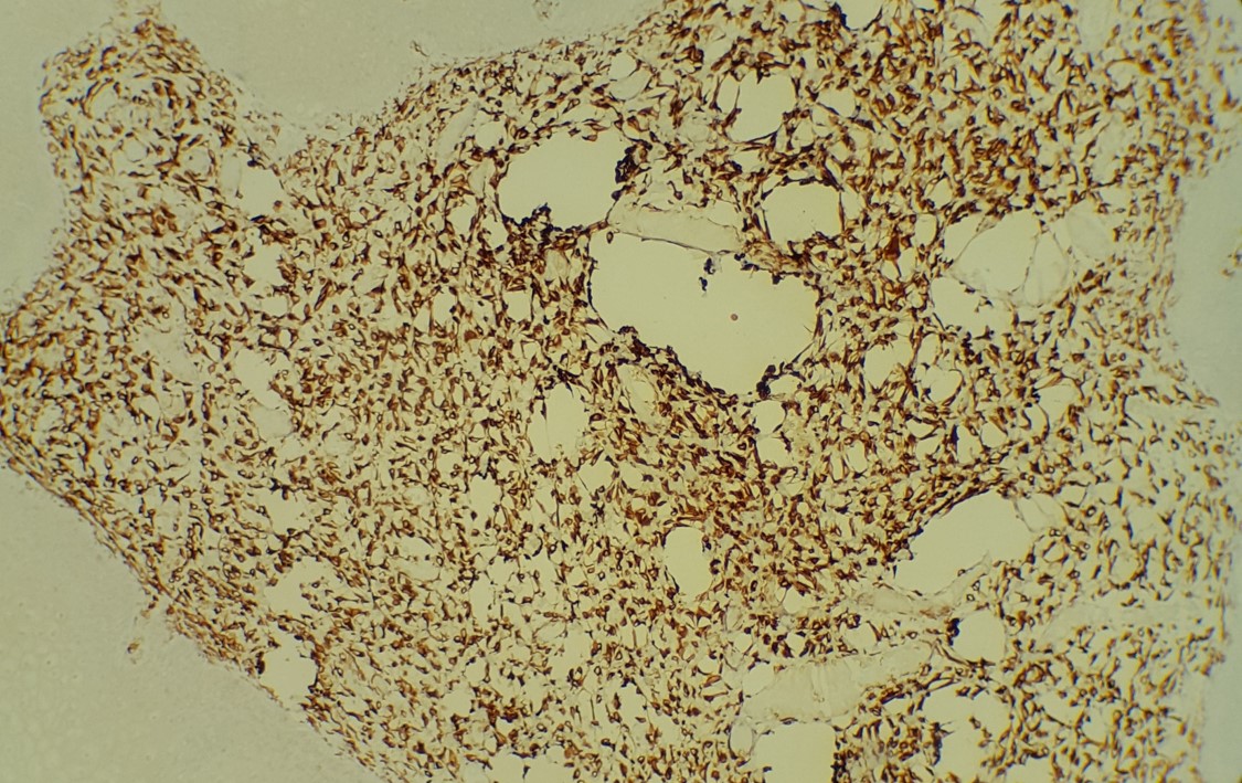
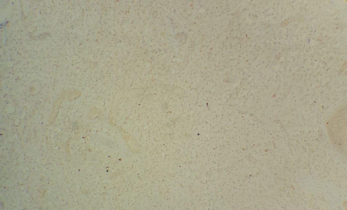
The findings in this case show a submucosal, nodular mass of bland spindle to oval cells and scattered mast cells in a background of thick collagen material. No atypia or mitotic figures are identified. The overlying mucosa is unremarkable. The immunoprofile of this lesion is CD34 and desmin positive, S-100 negative.
Superficial myofibroblastomas occur in adults and typically present as a superficial mass in the vagina, vulva, or cervix and average 2 to 3 cm in size. Microscopically, a moderately cellular proliferation of bland spindle cells are noted within collagenous stroma. Multiple patterns of growth can be present, including lacelike, fascicular, storiform, myxoid, and hyalinized, but a patternless pattern is most common. Thin-walled blood vessels can be seen concentrated in the center of the lesion. The immunoprofile shows positivity for ER, PR, desmin, vimentin staining and variable positivity for CD34. Keratins and S-100 are negative. Treatment is complete local excision as they have been reported to recur if incompletely excised.
Sources:
Soong, Thing R, Nucci, Marisa R, and Crum, Christopher P. “Neuroendocrine Carcinoma, Mixed Epithelial/Mesenchymal, and Mesenchymal Tumors, and Miscellaneous Lesions of the Cervix”. Diagnostic Gynecologic and Obstetric Pathology. 3rd ed. 2018.
Clement, Philip B and Young, Robert H. “Non-neoplastic Lesions and Benign Tumors of the Vulva”. Atlas of Gynecologic Surgical Pathology. 3rd ed. 2014.
Case contributed by: Joseph Drwiega, M.D., Fellow, Anatomic Pathology