Case History
A 59-year-old male presents with heavy proteinuria, hypoalbuminemia and normal kidney function. No other significant medical history. The findings of the kidney biopsy are classic for this entity. What antibody is associated with this diagnosis?
- PLA2R
- ANA
- ANCA
- Anti-GBM

The answer is “A” PLA2R.
Discussion:
The diagnosis is Membranous Glomerulopathy and should be suspected in all adults who present with unexplained proteinuria and hypoalbuminemia.
In addition to routine light microscopy, immunofluorescence and electron microscopy, staining for anti-phospholipase A2 receptor (PLA2R) antibodies by enzyme-linked immunosorbent assay [ELISA] should be performed in patients with characteristic histology of Membranous Glomerulopathy. Nearly all patients with positive staining for PLA2R have a diagnosis consistent with primary Membranous Glomerulopathy.
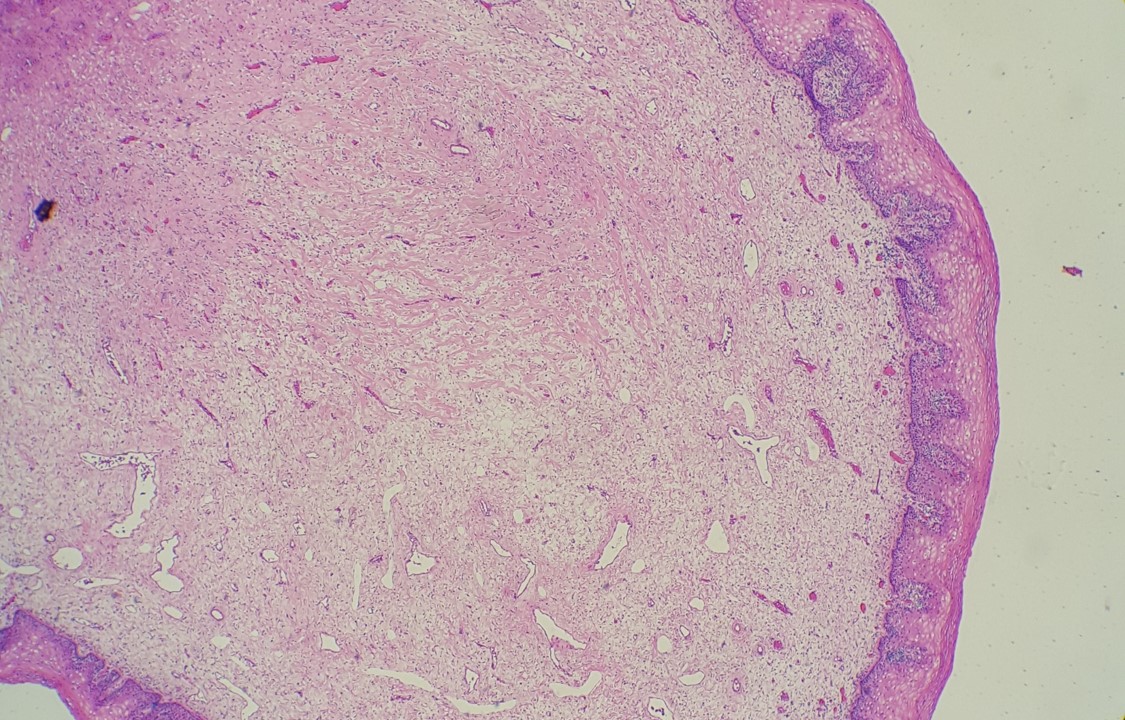
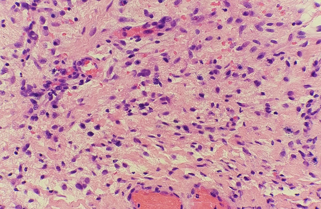
The typical histologic lesion on light microscopy is diffuse thickening of the glomerular basement membrane (GBM) throughout all glomeruli in the absence of significant hypercellularity. In some cases, "spikes" of GBM extending between the immune deposits may be seen with a silver stain.
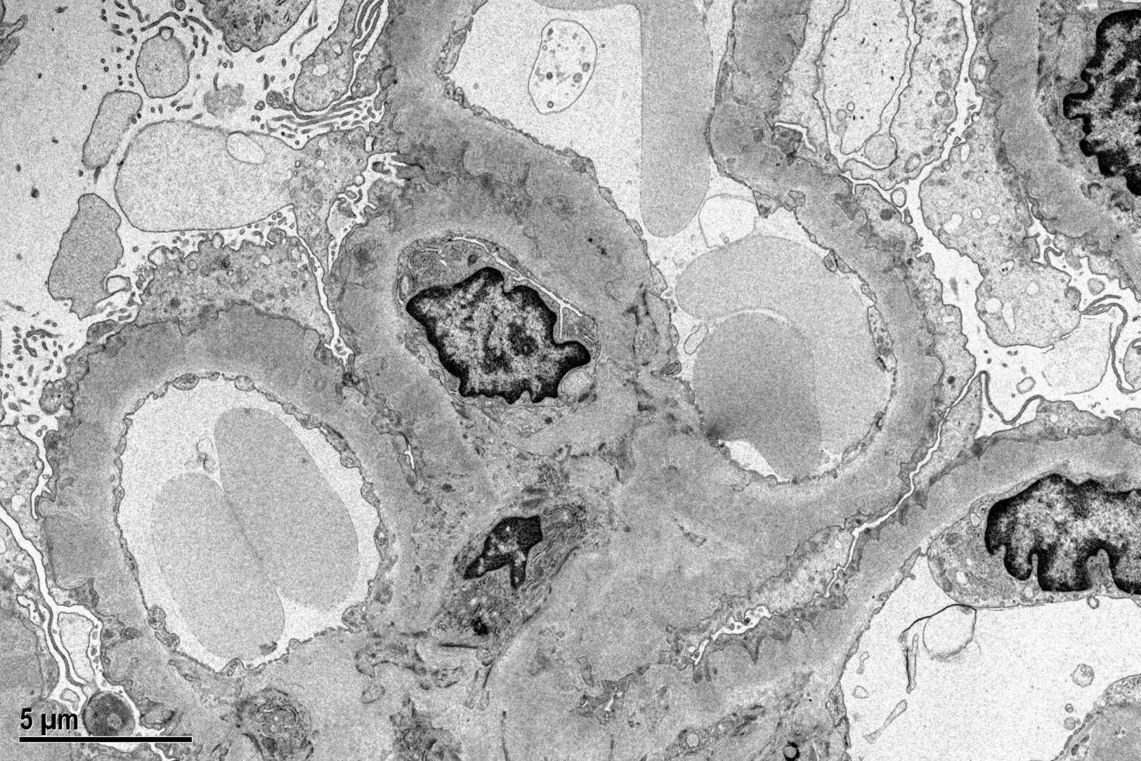
Immunofluorescence microscopy reveals a diffuse granular pattern of immunoglobulin G (IgG) staining along the GBM. The hallmark lesions on electron microscopy are subepithelial electron-dense deposits on the outer aspect of the GBM, effacement of the foot processes of the overlying podocytes, and expansion of the GBM by deposition of new extracellular matrix between the deposits (which are the "spikes" seen with the silver stain). Mesangial and/or subendothelial deposits of immunoglobulin are not seen in primary MN.
Case contributed by: Lindsay Matthews, M.D., Fellow, UAB Pathology