Case History
Patient history:
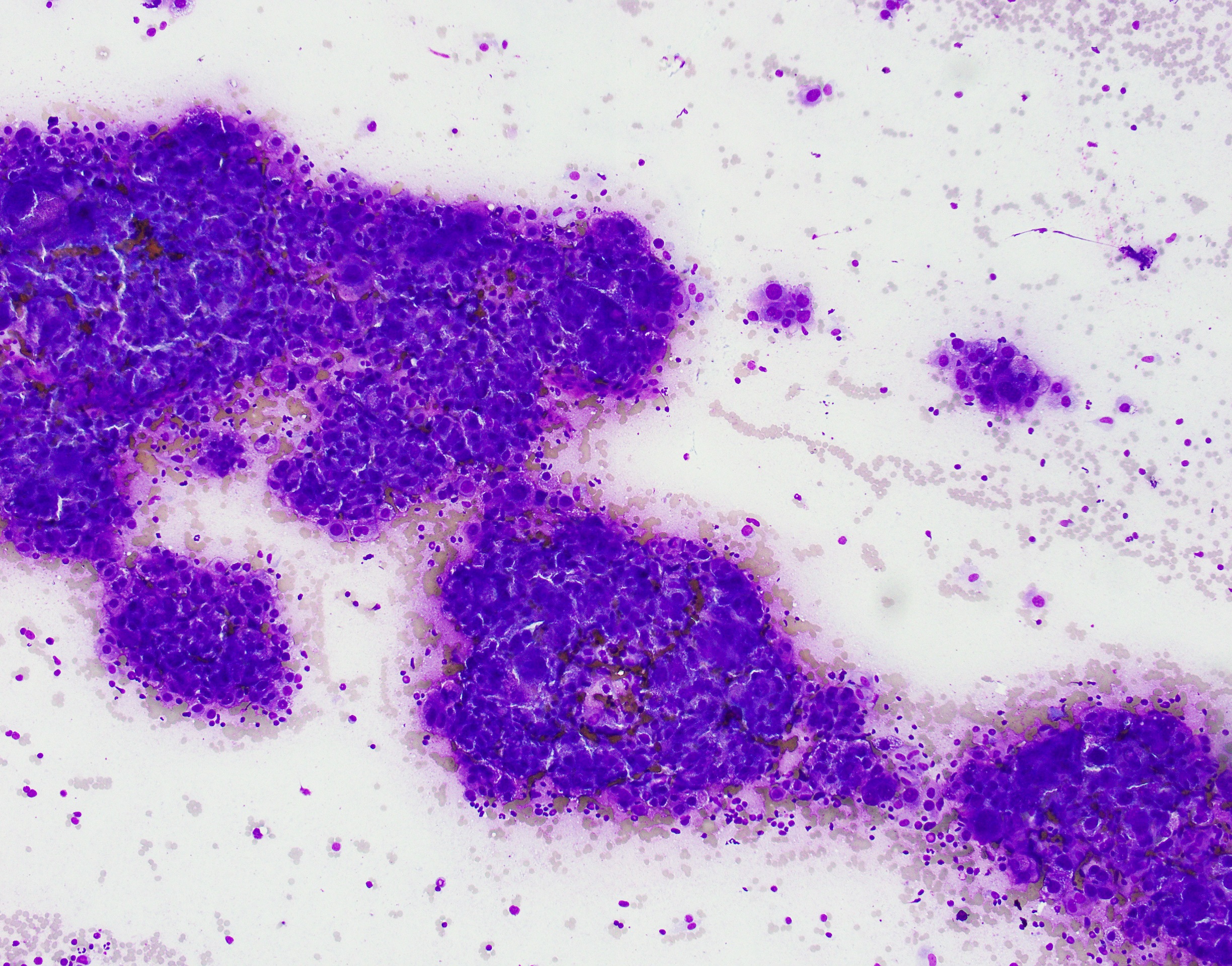
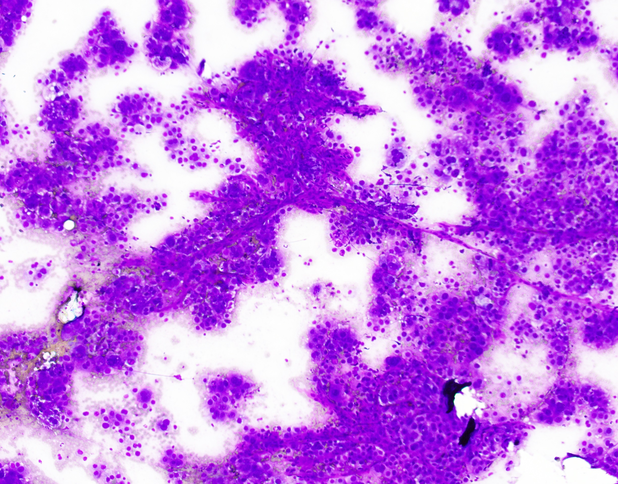
The patient is a 75-year-old male who was found to have multiple liver lesions and malignant appearing peri-pancreatic lymph nodes by radiology. Diff-Quik stained smears from the endoscopic FNA of the liver are shown.
What is the diagnosis?
- Metastatic clear cell renal cell carcinoma
- Metastatic ductal adenocarcinoma of the pancreas
- Hepatocellular carcinoma
- Epithelioid hemangioendothelioma

Answer: C. Hepatocellular carcinoma
Discussion:


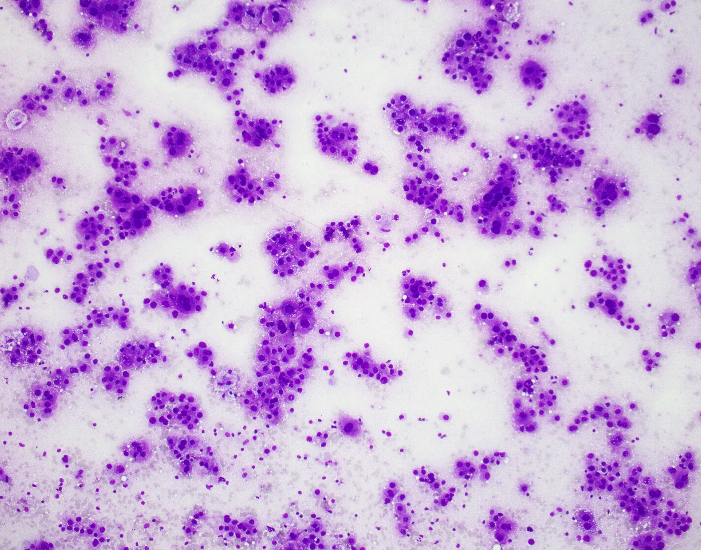
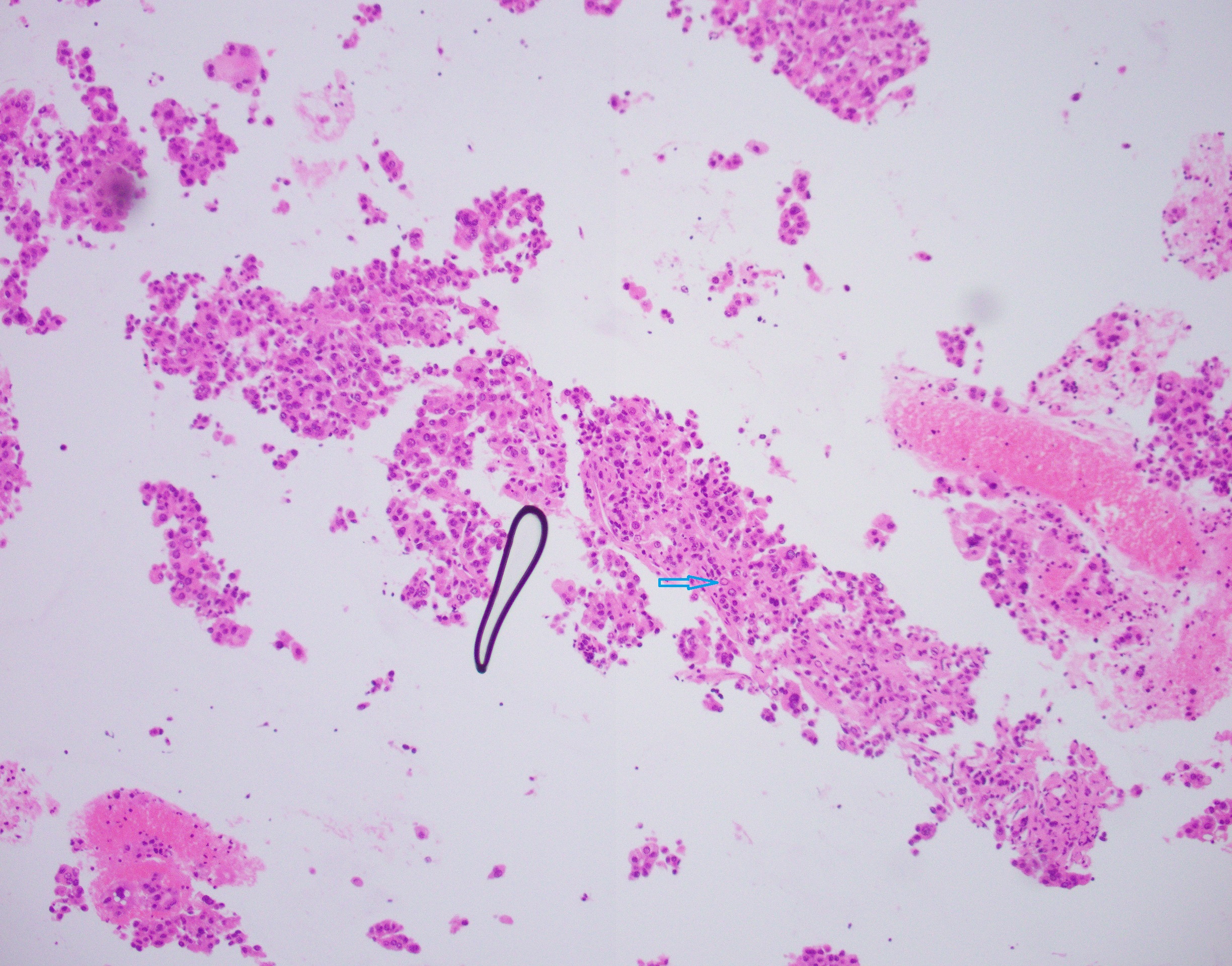
Hepatocellular carcinoma can present as a solitary nodule, multiple nodules, or as a diffuse liver enlargement. On cytology, hepatocytes are polygonal in shape with a centrally placed nucleus. Cytoplasmic bile can be noted as well. The smears for hepatocellular carcinoma (as the ones shown above) are usually highly cellular with isolated cells, high nuclear to cytoplasmic ratio, prominent nucleolus and large naked nuclei. Additional important cryptologic features include thickened cell cords with endothelial wrapping (Black arrow) and transgressing vessels (Red arrow) which are very characteristic. The features noted on cytology are in correlation with the H&E stained section from the cellblock, which also shows some intranuclear pseudoinclusions (Blue arrow).
Case contributed by: Morad Qarmali, M.D., Fellow, Cytopathology