Case History
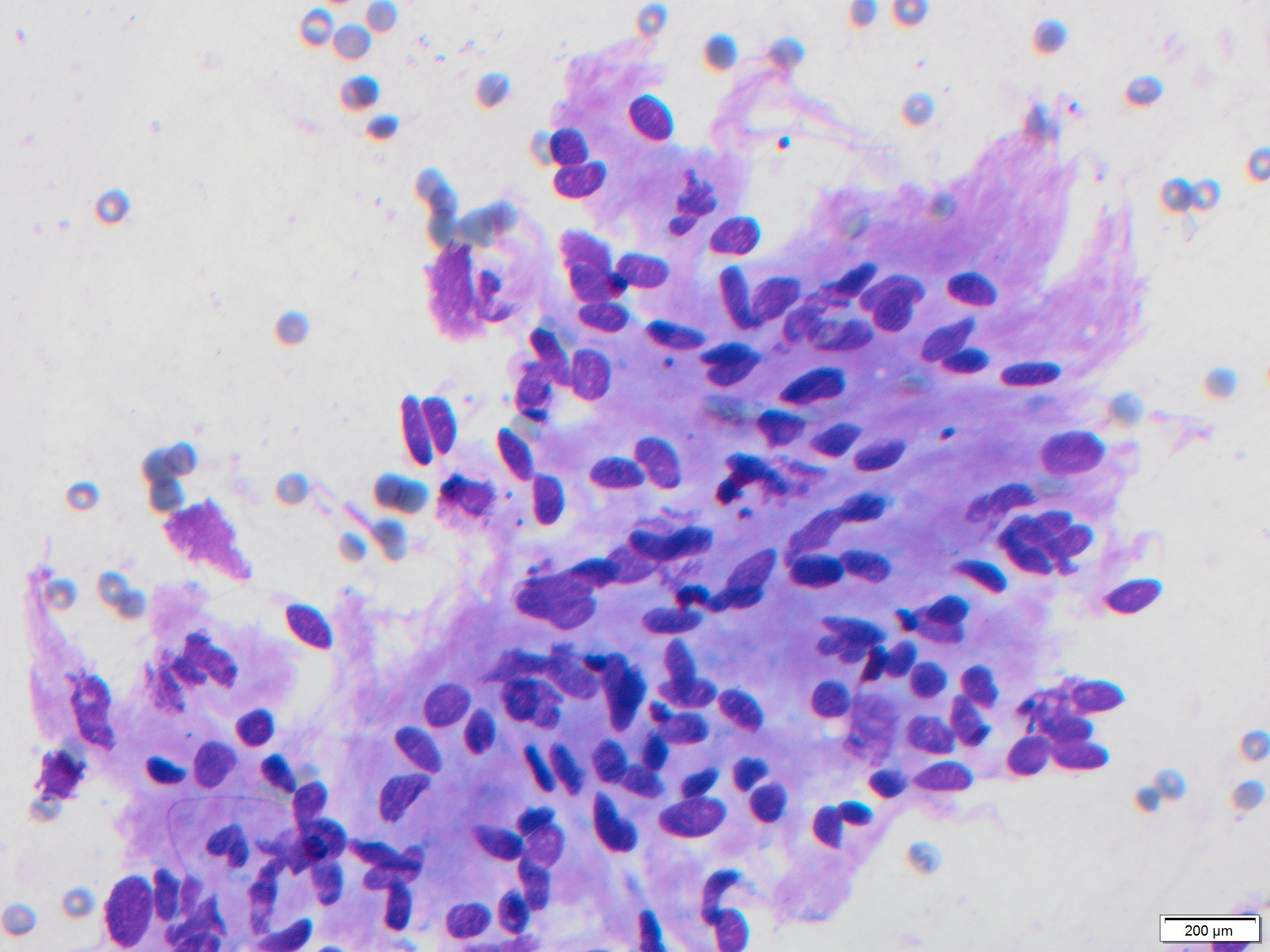
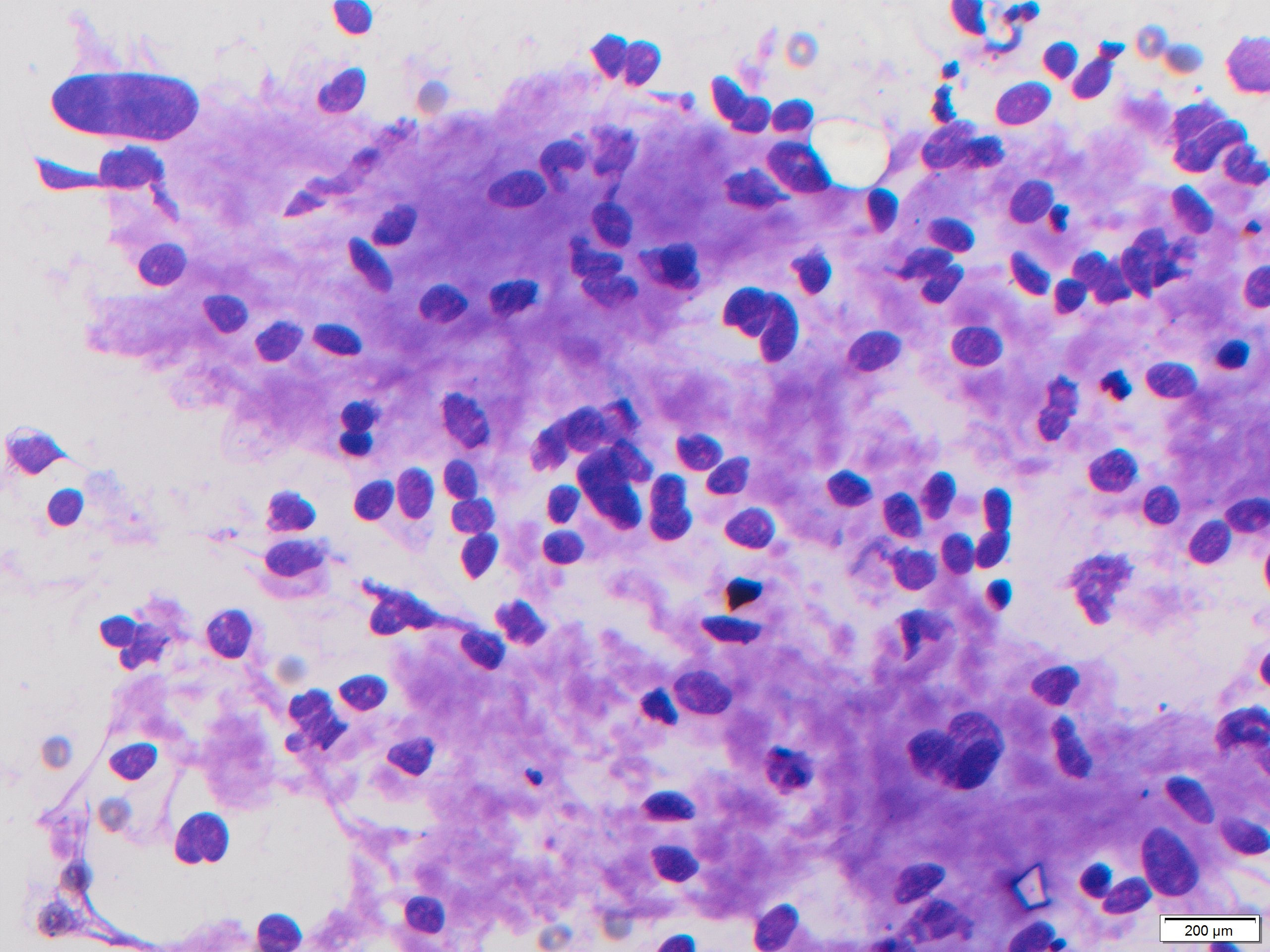
The patient is a 64-year-old man with a sublingual mass. Diff-Quik stained smears from fine needle aspirate are shown.
What is the diagnosis?
- Schwannoma
- Mucoepidermoid carcinoma
- Squamous cell carcinoma
- Pleomorphic adenoma
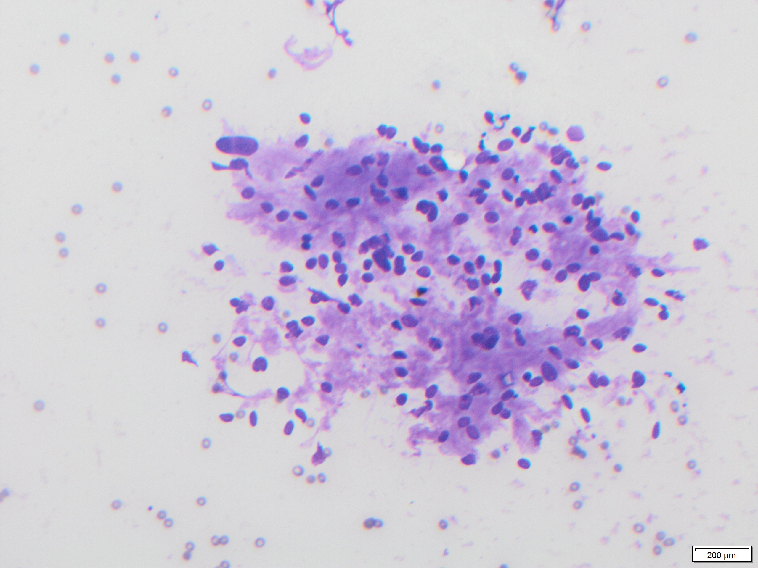
Answer: Schwannoma
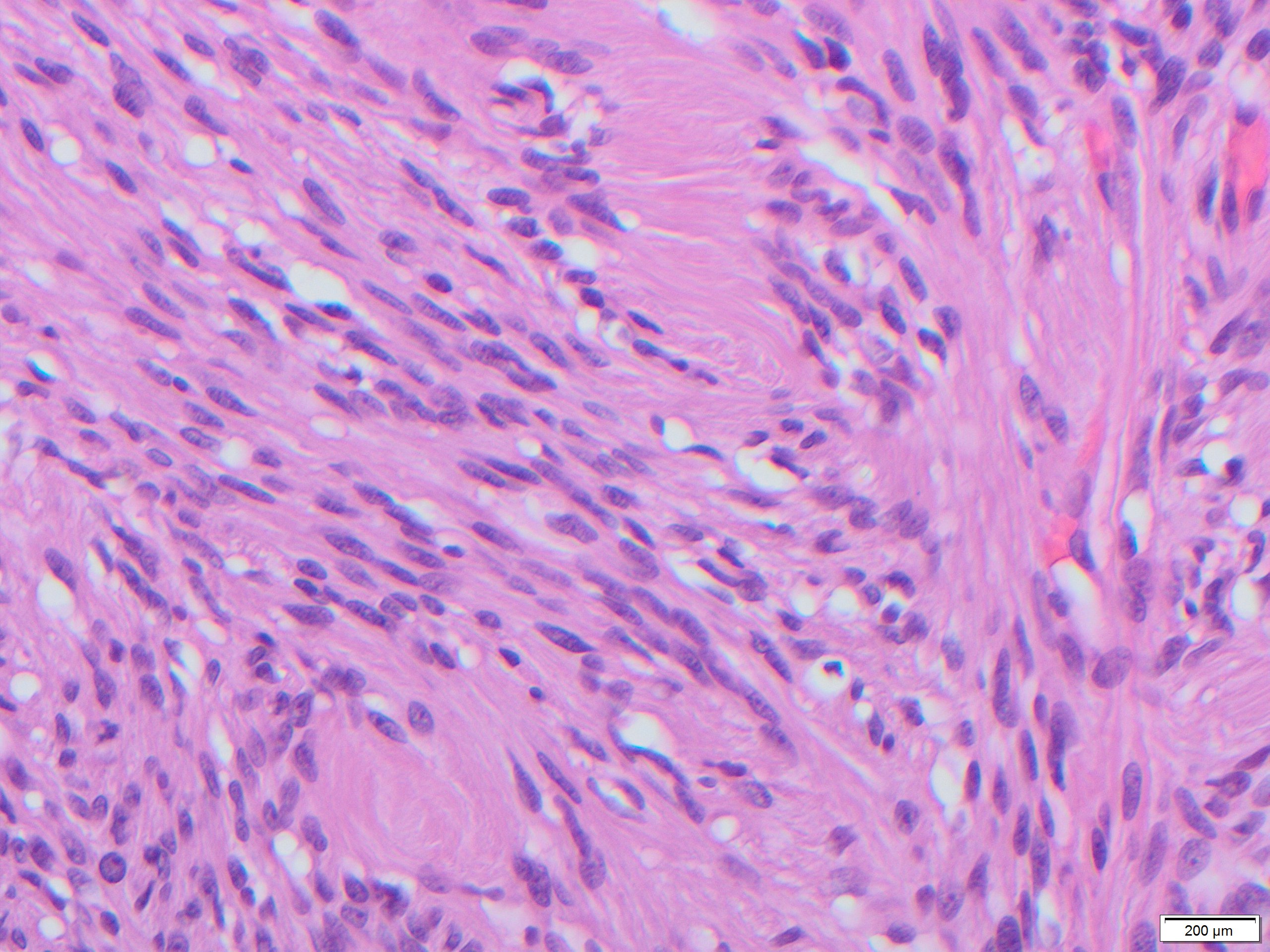
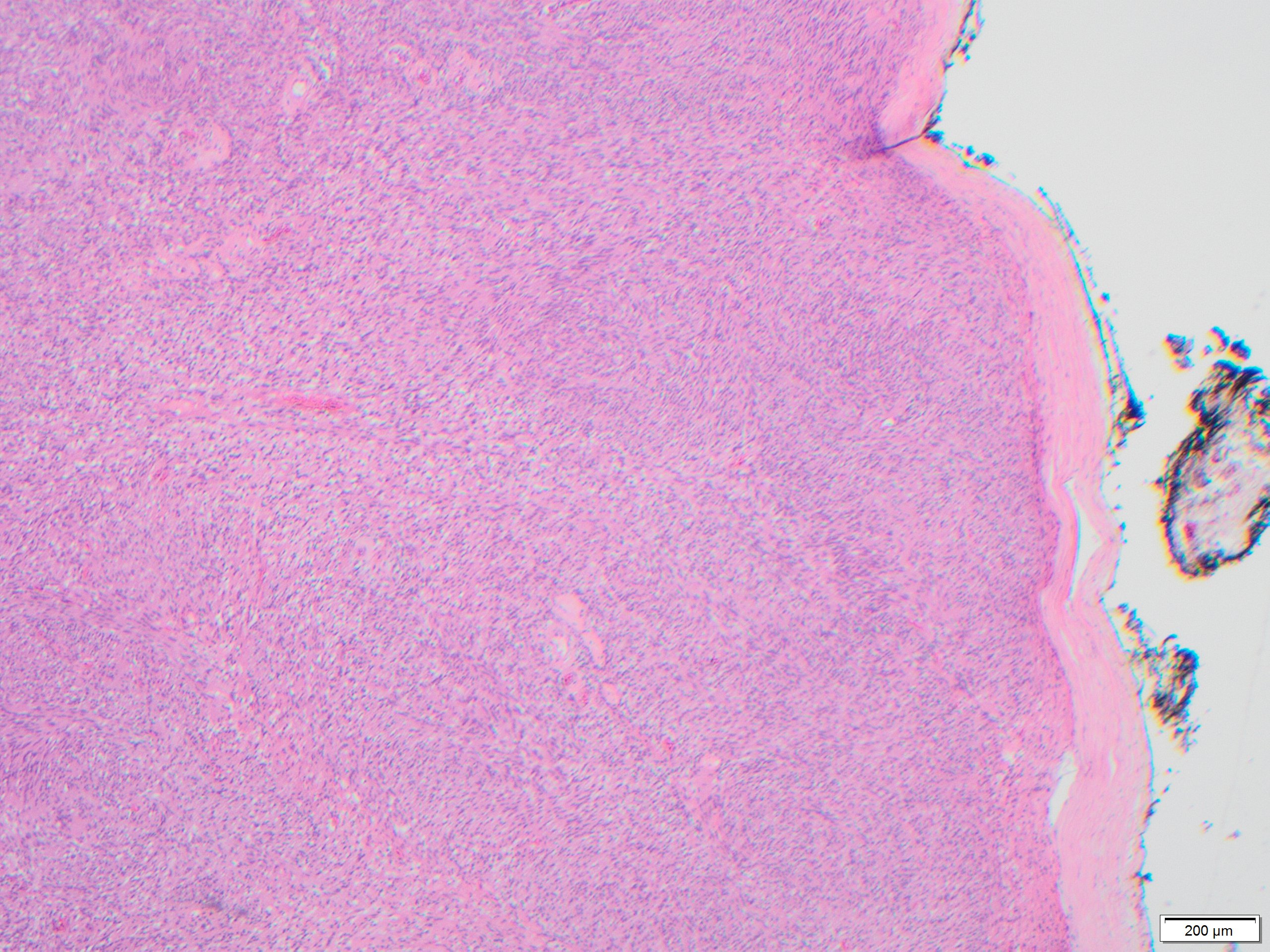
Nerve sheath tumors tend to be resistant to disaggregation on smears and therefore result in large syncytium-like fragments with irregular edges and very few to no singly-dispersed cells. The cells are elongated and spindled. The nuclei are oval, distinctively long and notched with tapered ends, and with minimal atypia. Palisades of nuclei alternating anucleate expanses of fibrillary cytoplasm (Verocay bodies) are present. Schwannomas often elicit pain during aspiration. Histologic sections show a well-circumscribed tumor with fibrous capsule, variable amounts of hypercellular Antoni A areas and rows of palisaded nuclei separated by fibrillary processes (Verocay bodies).
In this anatomic region, one cytomorphologic differential diagnosis would be pleomorphic adenoma of salivary gland, which would show two cell populations (ductal epithelial and myoepithelial) with matrix. The fibrillary matrix in pleomorphic adenoma is characteristically chondromyxoid, with sharply defined “troll-hair” fibers that have frayed edges, and most often stains bright magenta. Schwannoma has one cell population, and background material that is vaguely fibrillary and light pink, corresponding the cytoplasm of the cells. Additionally there is no salivary gland parenchyma in the background.
From the other options, Mucoepidermoid carcinoma would show epidermoid cells, mucocytes, intermediate cells and abundant mucin in background. Squamous cell carcinoma would display pronounced nuclear pleomorphism and atypia, keratin and keratinocytes.