Case History
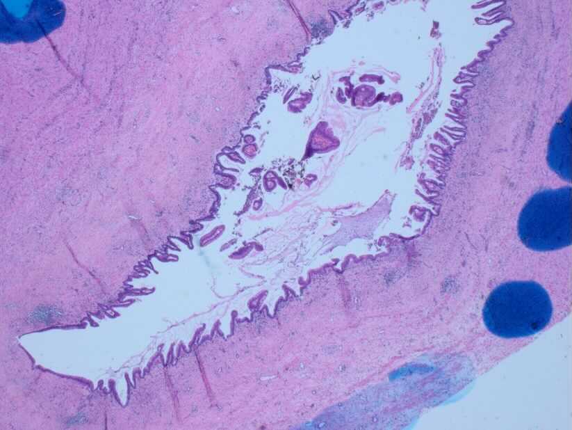
Case 1: 44 yr old female with appendicitis; appendix is grossly inflamed and dilated.

What is the diagnosis?
- LAMN
- HAMN
- Metastatic mucinous neoplasm
- Appendiceal diverticular disease
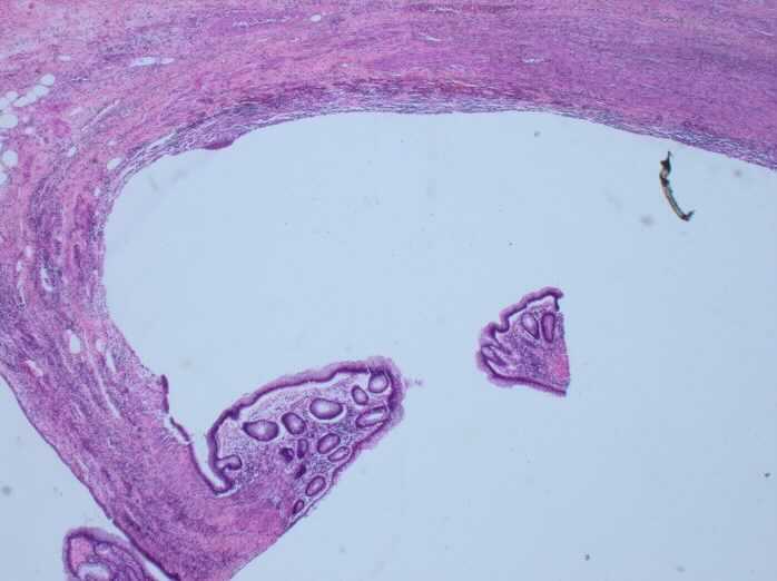
Case 2: 56 yr old female with a large dilated appendix; grossly a multiloculated cystic appendix.
What is the diagnosis?
- LAMN
- HAMN
- Metastatic mucinous neoplasm
- Appendiceal diverticular disease
Answers:
Case 1– Low-grade appendiceal mucinous neoplasm (LAMN)
Case 2- Appendiceal divertiucular disease (ADD)
Discussion:
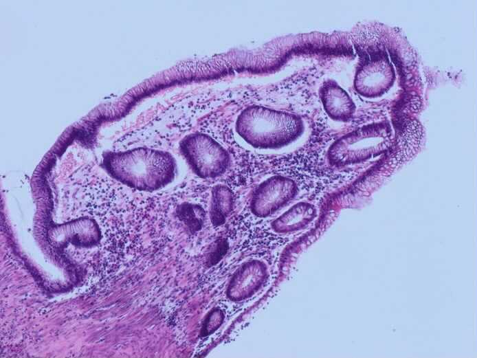
What are the key histological features that help in differentiation these entities? LAMN lacks muscularis mucosae and lamina propria. Fibrosis of the submucosa is frequent. ADD is often associated with attenuated epithelium; however, hyperplasia with mucus secretion may be seen. LAMN is often associated with KRAS and GNAS mutations. The two lesions, LAMN and ADD, may be seen within the same appendix.
Does LAMN often progress to high-grade appendiceal mucinous neoplasm (HAMN)? These are probably two different types of tumors. HAMN molecular pathway is also different than that of LAMN. As in other part of the intestine, does LAMN progress through stage Tis, to T1 to T2, T3 and T4? LAMN passes from Tis to T3 to T4. There is no stage T1 or T2 for LAMN.
Another important point: the number of neoplastic cells within extravasated mucin have prognostic value.