Case History
"A middle-aged male presented with liver mass. A biopsy of the liver lesion was negative for carcinoma. Clinical impression is consistent with cholangiocarcinoma with met to "periaortic lymph node". The patient went to OR. Enlarged periaortic lymph node was sent for frozen section.
What is the best answer?
A. Ectopic liver (EC)
B. Hepatocellular carcinoma (HCC)
C. Hepatic adenoma (HA)
D. ectopic liver (EL) with HA
Answer: D. Ectopic liver (EL) with HA
Discussion:
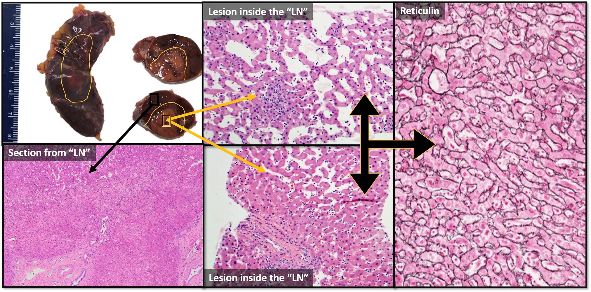
Ectopic liver (EL) is a rare developmental anomaly that has been reported in the abdominal cavity and rarely in the intrathoracic cavity. The most common location of ectopic liver is the gallbladder. EL can mimic malignant neoplasms radiologically and endoscopic excision is proven to be safe. In this case, we show ectopic liver located in abdominal peri-hepatic artery involved by incidental hepatocellular adenoma. On gross examination, the “lymph node” revealed benign hepatic parenchyma with a possible lesion (a paler area of hepatic parenchyma).
Microscopic examination of the lesion showed benign hepatic parenchyma in an abnormal location (ectopic liver) involved by hepatocellular adenoma. The adenoma demonstrates sinusoidal dilation, lack of portal structures, patchy inflammatory infiltrates grouped around vascular structures, minimal steatosis, and occasional interspersed thick-walled unpaired arteries. Reticulin special satin shows preserved meshwork, with typical 1 to 2 thickness in hepatocyte plates. No lymph node tissue was identified.